Image:PAM binding pocket correct.png
From Proteopedia
(Difference between revisions)
No higher resolution available.
PAM_binding_pocket_correct.png (640 × 304 pixel, file size: 94 KB, MIME type: image/png)
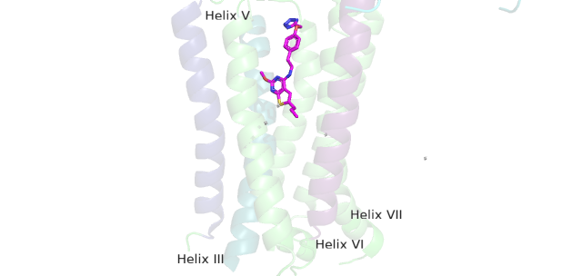
(Figure 3: This is PAM located in its binding pocket. PAM, JNJ-40411813, is shown in magenta and colored by atom. The image shows four labelled alpha helices (III, V, VI, and VII) that create the binding pocket in the 7TM region of mGlu2 for PAM to bind wi) |
|||
| Line 1: | Line 1: | ||
| - | + | This is PAM located in its binding pocket. | |
Current revision
This is PAM located in its binding pocket.
File history
Click on a date/time to view the file as it appeared at that time.
| Date/Time | User | Dimensions | File size | Comment | |
|---|---|---|---|---|---|
| (current) | 14:13, 22 March 2022 | Ashley R. Wilkinson (Talk | contribs) | 640×304 | 94 KB | Figure 3: This is PAM located in its binding pocket. PAM, JNJ-40411813, is shown in magenta and colored by atom. The image shows four labelled alpha helices (III, V, VI, and VII) that create the binding pocket in the 7TM region of mGlu2 for PAM to bind wi |
- Edit this file using an external application
See the setup instructions for more information.
Links
The following pages link to this file:
