We apologize for Proteopedia being slow to respond. For the past two years, a new implementation of Proteopedia has been being built. Soon, it will replace this 18-year old system. All existing content will be moved to the new system at a date that will be announced here.
3hf1
From Proteopedia
(Difference between revisions)
| Line 1: | Line 1: | ||
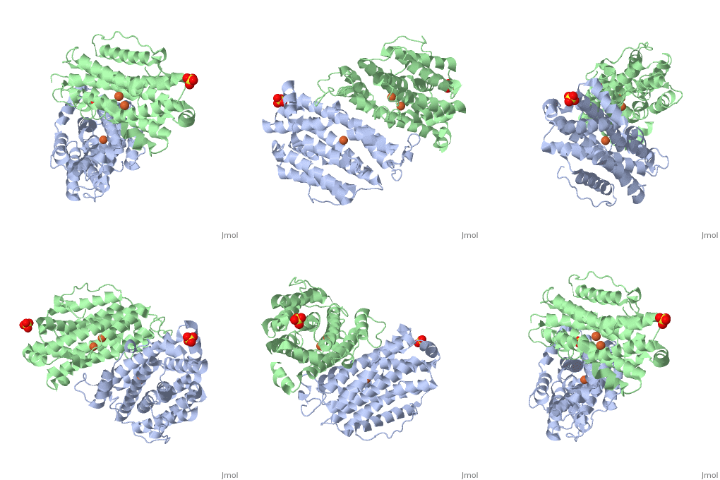
==Crystal structure of human p53R2== | ==Crystal structure of human p53R2== | ||
| - | <StructureSection load='3hf1' size='340' side='right' caption='[[3hf1]], [[Resolution|resolution]] 2.60Å' scene=''> | + | <StructureSection load='3hf1' size='340' side='right'caption='[[3hf1]], [[Resolution|resolution]] 2.60Å' scene=''> |
== Structural highlights == | == Structural highlights == | ||
| - | <table><tr><td colspan='2'>[[3hf1]] is a 2 chain structure with sequence from [ | + | <table><tr><td colspan='2'>[[3hf1]] is a 2 chain structure with sequence from [https://en.wikipedia.org/wiki/Human Human]. Full crystallographic information is available from [http://oca.weizmann.ac.il/oca-bin/ocashort?id=3HF1 OCA]. For a <b>guided tour on the structure components</b> use [https://proteopedia.org/fgij/fg.htm?mol=3HF1 FirstGlance]. <br> |
| - | </td></tr><tr id='ligand'><td class="sblockLbl"><b>[[Ligand|Ligands:]]</b></td><td class="sblockDat"><scene name='pdbligand=FE:FE+(III)+ION'>FE</scene>, <scene name='pdbligand=SO4:SULFATE+ION'>SO4</scene></td></tr> | + | </td></tr><tr id='ligand'><td class="sblockLbl"><b>[[Ligand|Ligands:]]</b></td><td class="sblockDat" id="ligandDat"><scene name='pdbligand=FE:FE+(III)+ION'>FE</scene>, <scene name='pdbligand=SO4:SULFATE+ION'>SO4</scene></td></tr> |
| - | <tr id='related'><td class="sblockLbl"><b>[[Related_structure|Related:]]</b></td><td class="sblockDat">[[1xsm|1xsm]], [[2uw2|2uw2]], [[1w69|1w69]], [[1w68|1w68]]</td></tr> | + | <tr id='related'><td class="sblockLbl"><b>[[Related_structure|Related:]]</b></td><td class="sblockDat"><div style='overflow: auto; max-height: 3em;'>[[1xsm|1xsm]], [[2uw2|2uw2]], [[1w69|1w69]], [[1w68|1w68]]</div></td></tr> |
| - | <tr id='gene'><td class="sblockLbl"><b>[[Gene|Gene:]]</b></td><td class="sblockDat">RRM2B, P53R2 ([ | + | <tr id='gene'><td class="sblockLbl"><b>[[Gene|Gene:]]</b></td><td class="sblockDat">RRM2B, P53R2 ([https://www.ncbi.nlm.nih.gov/Taxonomy/Browser/wwwtax.cgi?mode=Info&srchmode=5&id=9606 HUMAN])</td></tr> |
| - | <tr id='activity'><td class="sblockLbl"><b>Activity:</b></td><td class="sblockDat"><span class='plainlinks'>[ | + | <tr id='activity'><td class="sblockLbl"><b>Activity:</b></td><td class="sblockDat"><span class='plainlinks'>[https://en.wikipedia.org/wiki/Ribonucleoside-diphosphate_reductase Ribonucleoside-diphosphate reductase], with EC number [https://www.brenda-enzymes.info/php/result_flat.php4?ecno=1.17.4.1 1.17.4.1] </span></td></tr> |
| - | <tr id='resources'><td class="sblockLbl"><b>Resources:</b></td><td class="sblockDat"><span class='plainlinks'>[ | + | <tr id='resources'><td class="sblockLbl"><b>Resources:</b></td><td class="sblockDat"><span class='plainlinks'>[https://proteopedia.org/fgij/fg.htm?mol=3hf1 FirstGlance], [http://oca.weizmann.ac.il/oca-bin/ocaids?id=3hf1 OCA], [https://pdbe.org/3hf1 PDBe], [https://www.rcsb.org/pdb/explore.do?structureId=3hf1 RCSB], [https://www.ebi.ac.uk/pdbsum/3hf1 PDBsum], [https://prosat.h-its.org/prosat/prosatexe?pdbcode=3hf1 ProSAT]</span></td></tr> |
</table> | </table> | ||
== Disease == | == Disease == | ||
| - | [[ | + | [[https://www.uniprot.org/uniprot/RIR2B_HUMAN RIR2B_HUMAN]] Defects in RRM2B are the cause of mitochondrial DNA depletion syndrome type 8A (MTDPS8A) [MIM:[https://omim.org/entry/612075 612075]]. A disorder due to mitochondrial dysfunction characterized by various combinations of neonatal hypotonia, neurological deterioration, respiratory distress, lactic acidosis, and renal tubulopathy.<ref>PMID:17486094</ref> <ref>PMID:18504129</ref> Defects in RRM2B are the cause of mitochondrial DNA depletion syndrome type 8B (MTDPS8B) [MIM:[https://omim.org/entry/612075 612075]]. A disease due to mitochondrial dysfunction and characterized by ophthalmoplegia, ptosis, gastrointestinal dysmotility, cachexia, peripheral neuropathy. Defects in RRM2B are the cause of progressive external ophthalmoplegia with mitochondrial DNA deletions autosomal dominant type 5 (PEOA5) [MIM:[https://omim.org/entry/613077 613077]]. A disorder characterized by progressive weakness of ocular muscles and levator muscle of the upper eyelid. In a minority of cases, it is associated with skeletal myopathy, which predominantly involves axial or proximal muscles and which causes abnormal fatigability and even permanent muscle weakness. Ragged-red fibers and atrophy are found on muscle biopsy. A large proportion of chronic ophthalmoplegias are associated with other symptoms, leading to a multisystemic pattern of this disease. Additional symptoms are variable, and may include cataracts, hearing loss, sensory axonal neuropathy, ataxia, depression, hypogonadism, and parkinsonism.<ref>PMID:19664747</ref> |
== Function == | == Function == | ||
| - | [[ | + | [[https://www.uniprot.org/uniprot/RIR2B_HUMAN RIR2B_HUMAN]] Plays a pivotal role in cell survival by repairing damaged DNA in a p53/TP53-dependent manner. Supplies deoxyribonucleotides for DNA repair in cells arrested at G1 or G2. Contains an iron-tyrosyl free radical center required for catalysis. Forms an active ribonucleotide reductase (RNR) complex with RRM1 which is expressed both in resting and proliferating cells in response to DNA damage.<ref>PMID:10716435</ref> <ref>PMID:11517226</ref> <ref>PMID:11719458</ref> |
== Evolutionary Conservation == | == Evolutionary Conservation == | ||
[[Image:Consurf_key_small.gif|200px|right]] | [[Image:Consurf_key_small.gif|200px|right]] | ||
| Line 35: | Line 35: | ||
==See Also== | ==See Also== | ||
| - | + | *[[Ribonucleotide reductase 3D structures|Ribonucleotide reductase 3D structures]] | |
| - | *[[Ribonucleotide reductase|Ribonucleotide reductase]] | + | |
== References == | == References == | ||
<references/> | <references/> | ||
| Line 42: | Line 41: | ||
</StructureSection> | </StructureSection> | ||
[[Category: Human]] | [[Category: Human]] | ||
| + | [[Category: Large Structures]] | ||
[[Category: Ribonucleoside-diphosphate reductase]] | [[Category: Ribonucleoside-diphosphate reductase]] | ||
[[Category: Smith, P]] | [[Category: Smith, P]] | ||
Revision as of 12:46, 23 March 2022
Crystal structure of human p53R2
| |||||||||||
Categories: Human | Large Structures | Ribonucleoside-diphosphate reductase | Smith, P | Su, L | Tsai, S C | Yen, Y | Yuan, Y C | Zhou, B | Disease mutation | Dna damage | Dna repair | Dna replication | Iron | Metal-binding | Nucleus | Oxidoreductase | P53 inducible | Ribonucleotide reductase small subunit


