Sandbox Reserved 1716
From Proteopedia
(Difference between revisions)
| Line 33: | Line 33: | ||
==Vitamin K Epoxide== | ==Vitamin K Epoxide== | ||
| - | ==Warfarin== | ||
| - | <scene name='90/904321/Closedconformation/4'>Warfarin Binding</scene> | + | [[Image:Vitamin K epoxide.jpg|500 px|right|thumb|Figure 1. Vitamin K Epoxide structure]] |
| + | |||
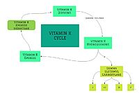
| + | As mentioned above, Vitamin K epoxide is a part of the Vitamin K cycle, necessary for blood coagulation. In the cycle, Vitamin K epoxide reductase (VKOR) reduces Vitamin K epoxide to quinone, or the active form of Vitamin K. What is occurring is VKOR donated electrons to Vitamin K epoxide, and those electrons come from the S-H of one of the cysteine pairs discussed above. The one cysteine pair has to be reduced for the transfer of electrons to the substrate can occur. | ||
| + | |||
| + | |||
| + | === Binding === | ||
| + | To start, VKOR is in its open conformation. The Vitamin K epoxide enters. The oxygens of the ketones bind to <scene name='90/904322/Tyr_asn_binding/1'>Asn80 and Tyr139</scene>. With Vitamin K epoxide in its place, the conformation of VKOR is partially oxidized in regards to the cysteine pairs, which overall leads to the reduction of the substrate. A disulfide bond forms between Cys51 and Cys132, resulting in the closed conformation. This leaves the sulfur on Cys43 and the sulfur on Cys135 protonated. The available hydrogens on these cysteines are utilized in reducing the epoxide. First, the sulfur on Cys51 and Cys43 form a new bond. The hydrogen from Cys43 binds to the oxygen in the epoxide. The sulfur on Cys132 and the sulfur on Cys15 then form a new disulfide bond. The hydrogen that was present on Cys135 forms a new bond with the oxygen of the epoxide. With these cysteine pairs formed, VKOR is left in an open conformation. The end products are the Vitamin K/quinone and water. | ||
| + | |||
| + | |||
| + | |||
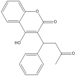
| + | == Warfarin == | ||
| + | |||
| + | [[Image:warfarin.jpg|400 px|righ | ||
| + | t|thumb|Figure 1. Warfarin]] | ||
| + | |||
| + | scene name='90/904321/Closedconformation/4'>Warfarin Binding</scene> | ||
</StructureSection> | </StructureSection> | ||
Revision as of 18:10, 27 March 2022
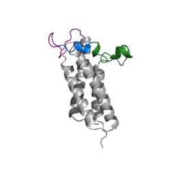
Vitamin K Epoxide Reductase
| |||||||||||
References
- ↑ Ransey E, Paredes E, Dey SK, Das SR, Heroux A, Macbeth MR. Crystal structure of the Entamoeba histolytica RNA lariat debranching enzyme EhDbr1 reveals a catalytic Zn(2+) /Mn(2+) heterobinucleation. FEBS Lett. 2017 Jul;591(13):2003-2010. doi: 10.1002/1873-3468.12677. Epub 2017, Jun 14. PMID:28504306 doi:http://dx.doi.org/10.1002/1873-3468.12677


