Sandbox Reserved 1716
From Proteopedia
(Difference between revisions)
| Line 10: | Line 10: | ||
== Introduction == | == Introduction == | ||
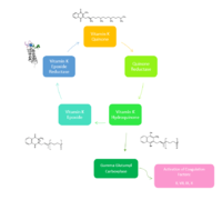
| - | [[Image:NewVitaminKCycle.PNG|200px|right|thumb|Figure 2. Overview of Vitamin K Cycle]] | + | [[Image:NewVitaminKCycle.PNG|200px|right|thumb|'''Figure 2. Overview of Vitamin K Cycle''': The cycle begins with Vitamin K Quinone. Vitamin K Quinone is reduced by enzyme Quinone Reductase. This leaves Vitamin K Hydroquinone which can either lead to Gamma Carboxylase activity that will activate Blood Coagulation Factors II, VII, IX, and X. After this, Vitamin K Epoxide is left over. Vitamin K Epoxide is reduced by the enzyme Vitamin K Epoxide Reductase to reform Vitamin K Quinone. ]] |
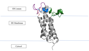
| - | '''Vitamin K Epoxide Reductase''' (VKOR) is an endoplasmic membrane enzyme that generates the active form of Vitamin K to support blood coagulation. VKOR homologs are known as integral membrane thiol oxidoreductases due to the function of VKOR being dependent on thiol residues and disulfide bonding. The vitamin K Cycle, and the VKOR enzyme specifically are common drug targets for thromboembolic diseases. This is because, as pictured, the vitamin K cycle is the process in which blood coagulant factors II, VII, IX, and X are activated. This promotes blood clotting, which (in extreme amounts) can be dangerous and cause thromboembolic diseases such as stroke, deep vein thrombosis, and/or pulmonary embolism. | + | '''Vitamin K Epoxide Reductase''' (VKOR) is an endoplasmic membrane enzyme that generates the active form of Vitamin K to support blood coagulation. VKOR homologs are known as integral membrane thiol oxidoreductases due to the function of VKOR being dependent on thiol residues and disulfide bonding. The vitamin K Cycle, and the VKOR enzyme specifically are common drug targets for thromboembolic diseases. This is because, as pictured, the vitamin K cycle is the process in which blood coagulant factors II, VII, IX, and X are activated. This promotes blood clotting, which (in extreme amounts) can be dangerous and cause thromboembolic diseases such as stroke, deep vein thrombosis, and/or pulmonary embolism. Vitamin K Epoxide Reductase is found and primarily synthesized in the liver. It is embedded in the membrane known as the endoplasmic reticulum. |
| + | |||
| + | == Structure == | ||
| + | <scene name='90/904321/Closedconformation/2'>Spinning VKOR</scene> | ||
| + | [https://en.wikipedia.org/wiki/Vitamin_K_epoxide_reductase VKOR WIKI] | ||
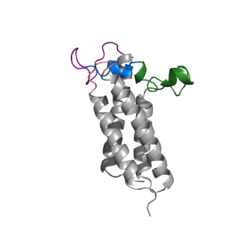
| + | The VKOR enzyme is made up of four transmembrane helices: T1, T2, T3, and T4.(Grey) Each of these helices come together to form a central pocket, that is topped by a cap domain. In the cap domain are important regions that are significant for Vitamin K binding, and the overall function of Vitamin K Epoxide Reductase. These important regions are the Anchor(Green), Cap Region (Blue), Beta Hairpin (Purple), and 3-4 Loop (Pink). The transmembrane helices form the central pocket that is also the active site of the enzyme. This is because the catalytic cysteines Cys132 and Cys135 are located in this region of the enzyme. | ||
| + | |||
| + | The transmembrane helices make up the ER-luminal region, which is large and flexible. Vitamin K Epoxide Reductase is known for its in-vitro instability. When trying to view the structure an extra protein known as sfGFP, superfolder green flourescent protein, is bound the N and C termini of Vitamin K Epoxide. For the purpose of viewing the structure, this protein has been removed from the pdb files. | ||
[[Image:VKORmembrane.png|300px|left|thumb|Figure 3. Orientation in Endoplasmic Reticulum]] | [[Image:VKORmembrane.png|300px|left|thumb|Figure 3. Orientation in Endoplasmic Reticulum]] | ||
| - | ===Location of Enzyme === | ||
| - | Vitamin K Epoxide Reductase is found and primarily synthesized in the liver. It is embedded in the membrane known as the endoplasmic reticulum. | ||
| Line 33: | Line 38: | ||
| - | == Structure == | ||
| - | <scene name='90/904321/Closedconformation/2'>Spinning VKOR</scene> | ||
| - | [https://en.wikipedia.org/wiki/Vitamin_K_epoxide_reductase VKOR WIKI] | ||
| - | The VKOR enzyme is made up of four transmembrane helices: T1, T2, T3, and T4.(Grey) Each of these helices come together to form a central pocket, that is topped by a cap domain. In the cap domain are important regions that are significant for Vitamin K binding, and the overall function of Vitamin K Epoxide Reductase. These important regions are the Anchor(Green), Cap Region (Blue), Beta Hairpin (Purple), and 3-4 Loop (Pink). The transmembrane helices form the central pocket that is also the active site of the enzyme. This is because the catalytic cysteines Cys132 and Cys135 are located in this region of the enzyme. | ||
| - | The transmembrane helices make up the ER-luminal region, which is large and flexible. Vitamin K Epoxide Reductase is known for its in-vitro instability. When trying to view the structure an extra protein known as sfGFP, superfolder green flourescent protein, is bound the N and C termini of Vitamin K Epoxide. For the purpose of viewing the structure, this protein has been removed from the pdb files. | ||
| - | + | ||
| + | |||
===Transmembrane Helices=== | ===Transmembrane Helices=== | ||
The Transmembrane helices are named Transmembrane Helix 1, Transmembrane Helix 2, Transmembrane Helix 3, and Transmembrane Helix 4. The residues on Transmembrane Helix 2 (TM2) and Transmembrane Helix 4 (TM4) are significant for the binding of Vitamin K to the hydrophobic pocket of the enzyme. Asparagine 83 on TM2 and Tyrosine 142 hydrogen bond to Vitamin K Epoxide, in order to hold it in place so that it may be reduced. The angle in which Vitamin K Epoxide binds is significant to the placement of the beta hairpin, and loop 3-4. Cysteine residues from the beta hairpin and loop 3-4 will donate their electrons to Vitamin K Epoxide to open the epoxide ring, and reform Vitamin K Quinone. | The Transmembrane helices are named Transmembrane Helix 1, Transmembrane Helix 2, Transmembrane Helix 3, and Transmembrane Helix 4. The residues on Transmembrane Helix 2 (TM2) and Transmembrane Helix 4 (TM4) are significant for the binding of Vitamin K to the hydrophobic pocket of the enzyme. Asparagine 83 on TM2 and Tyrosine 142 hydrogen bond to Vitamin K Epoxide, in order to hold it in place so that it may be reduced. The angle in which Vitamin K Epoxide binds is significant to the placement of the beta hairpin, and loop 3-4. Cysteine residues from the beta hairpin and loop 3-4 will donate their electrons to Vitamin K Epoxide to open the epoxide ring, and reform Vitamin K Quinone. | ||
Revision as of 17:15, 5 April 2022
Vitamin K Epoxide Reductase
| |||||||||||


