We apologize for Proteopedia being slow to respond. For the past two years, a new implementation of Proteopedia has been being built. Soon, it will replace this 18-year old system. All existing content will be moved to the new system at a date that will be announced here.
Sandbox Reserved 1703
From Proteopedia
(Difference between revisions)
| Line 19: | Line 19: | ||
===Inactive State=== | ===Inactive State=== | ||
| - | A few hallmarks of the inactive structure of mGlu2 are the <scene name='90/904307/Better_inactive_structure/3'>Venus Fly Trap Domain (VFT)</scene> in the open conformation, well separated <scene name='90/904307/Better_inactive_structure/2'>Cysteine Rich Domain</scene>, and distinct orientation of the 7 Transmembrane Domains (7TM). The most critical component of the inactive form is the <scene name='90/904307/Tmd_helices/4' | + | A few hallmarks of the inactive structure of mGlu2 are the <scene name='90/904307/Better_inactive_structure/3'>Venus Fly Trap Domain (VFT)</scene> in the open conformation, well separated <scene name='90/904307/Better_inactive_structure/2'>Cysteine Rich Domain</scene>, and distinct orientation of the 7 Transmembrane Domains (7TM). The most critical component of the inactive form is the <scene name='90/904307/Tmd_helices/4'>asymmetric TM3-TM4 interface</scene> formed by the 7 α-helices in the α and β chains of the 7TM. The inactive structure of mGlu2 is mediated mainly by helices 3 and 4 on both the α and β chains of the dimer through hydrophobic interactions. These <scene name='90/904307/Tm3-tm4_hydrophobic/2'>hydrophobic interactions</scene> between both transmembrane helices stabilize inactive conformation of mGlu2<ref name="Lin"/>. Key hydrophobic interactions are between A630 V699 on the helix |
===Intermediate Form=== | ===Intermediate Form=== | ||
| Line 25: | Line 25: | ||
===PAM and NAM Bound Form=== | ===PAM and NAM Bound Form=== | ||
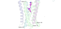
| - | A positive allosteric modulator (PAM) or a negative allosteric modulator (NAM) can bind to mGlu2. PAM and NAM induce different conformational changes, which result in different outcomes. <scene name='90/904308/Pam/3'>PAM binds</scene> to the receptor, induces conformational changes, which helps to promote greater affinity for G protein binding. PAM binds in a binding pocket that is created by helices III, V, VI, VII in the TMD. Within helix VI, the hydrophobic is is composed of W773, F776, L777, and F780. Due to spatial hindrance, helix VI is shifted downward, causing conformational changes. NAM, however, reduces the affinity for G protein binding. NAM binds to the same binding pocket as PAM and also interacts with residue W773, but NAM occupies the binding site a little deeper than PAM. This causes NAM to push the side chain of W773 towards helix VII<ref name="Lin"/>. | + | A positive allosteric modulator (PAM) or a negative allosteric modulator (NAM) can bind to mGlu2. PAM and NAM induce different conformational changes, which result in different outcomes. <scene name='90/904308/Pam/3'>PAM binds</scene> to the receptor, induces conformational changes, which helps to promote greater affinity for G protein binding. PAM binds in a binding pocket that is created by helices III, V, VI, VII in the <scene name='90/904307/Tmd_helices/4'>TMD</scene>. Within helix VI, the hydrophobic is is composed of W773, F776, L777, and F780. Due to spatial hindrance, helix VI is shifted downward, causing conformational changes. NAM, however, reduces the affinity for G protein binding. NAM binds to the same binding pocket as PAM and also interacts with residue W773, but NAM occupies the binding site a little deeper than PAM. This causes NAM to push the side chain of W773 towards helix VII<ref name="Lin"/>. |
[[Image:PAM binding pocket correct.png |250px|right|thumb|'''Figure 4.'''PAM located binding pocket. PAM, JNJ-40411813, is shown in magenta and colored by atom type, four labelled binding helices (III, V, VI, and VII) create the binding pocket in the 7TM region for PAM binding. PAM binding promotes G-protein activation by mGLu2.]] | [[Image:PAM binding pocket correct.png |250px|right|thumb|'''Figure 4.'''PAM located binding pocket. PAM, JNJ-40411813, is shown in magenta and colored by atom type, four labelled binding helices (III, V, VI, and VII) create the binding pocket in the 7TM region for PAM binding. PAM binding promotes G-protein activation by mGLu2.]] | ||
Revision as of 23:46, 14 April 2022
Metabotropic Glutamate Receptor 2
| |||||||||||
References
- ↑ 1.0 1.1 1.2 1.3 1.4 1.5 1.6 1.7 1.8 Lin S, Han S, Cai X, Tan Q, Zhou K, Wang D, Wang X, Du J, Yi C, Chu X, Dai A, Zhou Y, Chen Y, Zhou Y, Liu H, Liu J, Yang D, Wang MW, Zhao Q, Wu B. Structures of Gi-bound metabotropic glutamate receptors mGlu2 and mGlu4. Nature. 2021 Jun;594(7864):583-588. doi: 10.1038/s41586-021-03495-2. Epub 2021, Jun 16. PMID:34135510 doi:http://dx.doi.org/10.1038/s41586-021-03495-2
- ↑ 2.0 2.1 Seven, Alpay B., et al. “G-Protein Activation by a Metabotropic Glutamate Receptor.” Nature News, Nature Publishing Group, 30 June 2021, https://www.nature.com/articles/s1586-021-03680-3
- ↑ Du, Juan, et al. “Structures of Human mglu2 and mglu7 Homo- and Heterodimers.” Nature News, Nature Publishing Group, 16 June 2021, https://www.nature.com/articles/s41586-021-03641-w.>
- ↑ 4.0 4.1 4.2 4.3 Muguruza C, Meana JJ, Callado LF. Group II Metabotropic Glutamate Receptors as Targets for Novel Antipsychotic Drugs. Front Pharmacol. 2016 May 20;7:130. doi: 10.3389/fphar.2016.00130. eCollection, 2016. PMID:27242534 doi:http://dx.doi.org/10.3389/fphar.2016.00130
- ↑ Ellaithy A, Younkin J, Gonzalez-Maeso J, Logothetis DE. Positive allosteric modulators of metabotropic glutamate 2 receptors in schizophrenia treatment. Trends Neurosci. 2015 Aug;38(8):506-16. doi: 10.1016/j.tins.2015.06.002. Epub, 2015 Jul 4. PMID:26148747 doi:http://dx.doi.org/10.1016/j.tins.2015.06.002
Student Contributors
Frannie Brewer and Ashley Wilkinson


