We apologize for Proteopedia being slow to respond. For the past two years, a new implementation of Proteopedia has been being built. Soon, it will replace this 18-year old system. All existing content will be moved to the new system at a date that will be announced here.
Sandbox Reserved 1716
From Proteopedia
(Difference between revisions)
| Line 13: | Line 13: | ||
<scene name='90/904321/Vitamin_k_epoxide_reductase/1'>Vitamin K Epoxide Reductase</scene> | <scene name='90/904321/Vitamin_k_epoxide_reductase/1'>Vitamin K Epoxide Reductase</scene> | ||
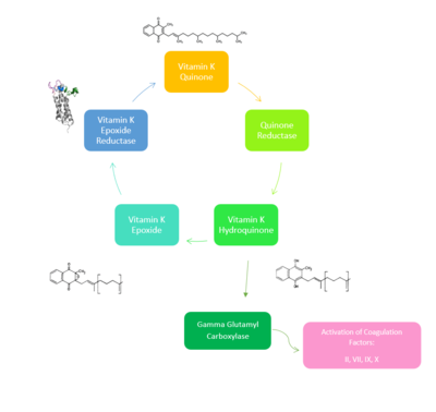
[https://en.wikipedia.org/wiki/Vitamin_K_epoxide_reductase VKOR WIKI](VKOR) is an endoplasmic membrane enzyme that generates the active form of Vitamin K to support blood coagulation. VKOR homologs are integral membrane thiol oxidoreductases [https://en.wikipedia.org/wiki/Thiol_oxidoreductase Thiol OxidoReductase] due to the function of VKOR being dependent on thiol residues and disulfide bonding. The Vitamin K Cycle and the VKOR enzyme specifically are common drug targets for thromboembolic diseases. This is because, as pictured, the vitamin K cycle is required to activate blood coagulant factors [https://en.wikipedia.org/wiki/Thrombin II], [https://en.wikipedia.org/wiki/Coagulation_factor_VII VII], [https://en.wikipedia.org/wiki/Factor_IX IX], and [https://en.wikipedia.org/wiki/Factor_X#:~:text=Factor%20X%2C%20also%20known%20by,vitamin%20K%20for%20its%20synthesis. X]. Coagulant factor activation promotes blood clotting, which in high amounts can be dangerous and cause thromboembolic diseases such as stroke, deep vein thrombosis, and/or pulmonary embolism. | [https://en.wikipedia.org/wiki/Vitamin_K_epoxide_reductase VKOR WIKI](VKOR) is an endoplasmic membrane enzyme that generates the active form of Vitamin K to support blood coagulation. VKOR homologs are integral membrane thiol oxidoreductases [https://en.wikipedia.org/wiki/Thiol_oxidoreductase Thiol OxidoReductase] due to the function of VKOR being dependent on thiol residues and disulfide bonding. The Vitamin K Cycle and the VKOR enzyme specifically are common drug targets for thromboembolic diseases. This is because, as pictured, the vitamin K cycle is required to activate blood coagulant factors [https://en.wikipedia.org/wiki/Thrombin II], [https://en.wikipedia.org/wiki/Coagulation_factor_VII VII], [https://en.wikipedia.org/wiki/Factor_IX IX], and [https://en.wikipedia.org/wiki/Factor_X#:~:text=Factor%20X%2C%20also%20known%20by,vitamin%20K%20for%20its%20synthesis. X]. Coagulant factor activation promotes blood clotting, which in high amounts can be dangerous and cause thromboembolic diseases such as stroke, deep vein thrombosis, and/or pulmonary embolism. | ||
| - | |||
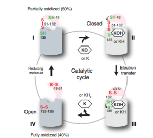
Vitamin K Epoxide Reductase is found and primarily synthesized in the liver. In the liver, the VKOR enzyme is set in the endoplasmic reticulum membrane (Fig.2). The transmembrane helices are located in the Endoplasmic Reticulum Luminal Region, which is the region between the ER Lumen and the Cytosol. The cap region is partially oriented in the ER Lumen. The active site remains within the ER membrane.The Anchor is partially within the ER lumen, and partially embedded in the ER membrane. The anchor is what attaches the cap domain and stabilizes it, which allows the cap domain to cover the active site. Vitamin K Epoxide Reductase is unstable in-vitro. To determine its structure an extra protein superfolder green flourescent protein was appended to the N and C termini of Vitamin K Epoxide. For the visualizing VKOR, this protein has been removed from the structural scenes. After superfolder green flourescent protein was removed from the structural scenes, we further took the structure files and resequenced them to better align with the numbering of the protein. In these files the sequence slightly differs between the organisms used to view Vitamin K Epoxide Reductase. In the human version, HsVKOR, the catalytic cysteines that play an intricate role in the reduction of Vitamin K Epoxide are cysteines 43, 51, 132, and 135. In the pufferfish version of the file, TrVKORL, the cysteines are 52, 55, 141, and 144. | Vitamin K Epoxide Reductase is found and primarily synthesized in the liver. In the liver, the VKOR enzyme is set in the endoplasmic reticulum membrane (Fig.2). The transmembrane helices are located in the Endoplasmic Reticulum Luminal Region, which is the region between the ER Lumen and the Cytosol. The cap region is partially oriented in the ER Lumen. The active site remains within the ER membrane.The Anchor is partially within the ER lumen, and partially embedded in the ER membrane. The anchor is what attaches the cap domain and stabilizes it, which allows the cap domain to cover the active site. Vitamin K Epoxide Reductase is unstable in-vitro. To determine its structure an extra protein superfolder green flourescent protein was appended to the N and C termini of Vitamin K Epoxide. For the visualizing VKOR, this protein has been removed from the structural scenes. After superfolder green flourescent protein was removed from the structural scenes, we further took the structure files and resequenced them to better align with the numbering of the protein. In these files the sequence slightly differs between the organisms used to view Vitamin K Epoxide Reductase. In the human version, HsVKOR, the catalytic cysteines that play an intricate role in the reduction of Vitamin K Epoxide are cysteines 43, 51, 132, and 135. In the pufferfish version of the file, TrVKORL, the cysteines are 52, 55, 141, and 144. | ||
| - | + | [[Image:VKORcycle.PNG|300px|right|thumb|Figure 2: The catalytic cycle of Vitamin K Epoxide Reductase <ref name=”Shixuan”>PMID:33154105</ref> ]] | |
| - | + | ||
| - | + | ||
| - | + | ||
| - | + | ||
| - | + | ||
| - | + | ||
| - | + | ||
| - | + | ||
| - | + | ||
| - | + | ||
| - | + | ||
| - | + | ||
| - | [[Image:VKORcycle.PNG|300px|right|thumb|Figure | + | |
| - | + | ||
| - | + | ||
| - | + | ||
| - | + | ||
| - | + | ||
| Line 62: | Line 43: | ||
| + | ==Step | ||
| - | The transmembrane helices are located in the Endoplasmic Reticulum Luminal Region, which is the region between the ER Lumen and the Cytosol. Vitamin K Epoxide Reductase is unstable in-vitro. To determine its structure an extra protein, sfGFP: superfolder green flourescent protein, was appended to the N and C termini of Vitamin K Epoxide. For the visualizing VKOR, this protein has been removed from the structural scenes. | ||
| - | |||
| - | |||
| - | ===Transmembrane Helices=== | ||
| - | The Transmembrane region has four helices, labeled 1-4. Transmembrane Helix 2 (TM2) and 4 (TM4) contribute to the binding of Vitamin K to the hydrophobic pocket for reduction. Asparagine 83 on TM2 and Tyrosine 142 on TM4 hydrogen bond to Vitamin K Epoxide to hold it in the correct orientation for reduction. The angle in which Vitamin K Epoxide binds in the central pocket is significant to the placement of the beta hairpin, and loop 3-4, Cysteine residues C51-C132 will donate their electrons to Vitamin K Epoxide (Fig. 4) to open the epoxide ring, and reform Vitamin K Quinone (Fig. 2). | ||
| - | |||
| - | ===Cap Domain=== | ||
| - | |||
| - | The cap domain of Vitamin K Epoxide Reductase plays an intricate role in its function. When Vitamin K Epoxide (or a similar substrate) binds in the hydrophobic pocket of VKOR, the cap domain undergoes a conformational change that will allow for specific cysteine residues to be able to open the epoxide ring and to recreate Vitamin K Quinone. The catalytic cycle begins in an open fully oxidized conformation. <scene name='90/904321/Openvkor/3'>Open Oxidized Conformation</scene> This conformation has slightly different parts. These include the Anchor (green), the cap region (blue), 3-4 Loop (pink), and luminal helix (yellow). When Vitamin K Epoxide binds, the entire cap domain undergoes a slight conformation change, but the luminal helix has a larger change. The luminal helix (yellow) bends forward where specific cysteines on this region are in proximity to other important cysteines. The luminal helix is then referred to as the beta hairpin (purple). <scene name='90/904321/Vkoclosed/3'>Partially Oxidized Closed Conformation</scene> | ||
| - | |||
| - | =Catalytic Cysteines= | ||
| - | A set of four catalytic are required for Vitamin K Epoxide reduction and are consistently conserved in all VKOR homologs. In the human homolog (HsVKOR) these cysteines are Cys43, Cys51, Cys132, and Cys 135. <scene name='90/904321/Cysteines/6'>Significant Cysteines</scene> In the Pufferfish homolog (TrVKORL) these cysteines, due to Cryo-EM differences,are Cys52, Cys55, Cys141, and Cys144. These cysteines donate their electrons to the epoxide ring on Vitamin K Epoxide which reforms Vitamin K Quinone. In the closed active conformation, that is induced when Vitamin K binds in the hydrophobic pocket. (NEED TO FINISH) | ||
Revision as of 14:20, 15 April 2022
Vitamin K Epoxide Reductase
| |||||||||||
