Sandbox Reserved 1715
From Proteopedia
(Difference between revisions)
| Line 37: | Line 37: | ||
[[Image: Active vs inactive vft.png|250px|left|thumb|Figure 2. Cysteine 121 positioning in the inactive (left) vs the active (right) mGlu conformation ]] | [[Image: Active vs inactive vft.png|250px|left|thumb|Figure 2. Cysteine 121 positioning in the inactive (left) vs the active (right) mGlu conformation ]] | ||
==== Domains ==== | ==== Domains ==== | ||
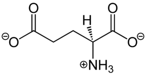
| - | <scene name='90/904320/Mglu2_domains_vft/3'>VFT</scene>: The extracellular location in which the two glutamate agonists bind is known as the VFT. This domain includes a disulfide bond between C121 of the alpha and beta chains. This bond is shifted down in the inactive form and undergoes an upward movement upon glutamate binding which stabilizes the <scene name='90/904319/Vft/1'>active site</scene> (Figure 2) Glutamate binds within the <scene name='90/904320/Active_site_interactions/3'> | + | <scene name='90/904320/Mglu2_domains_vft/3'>VFT</scene>: The extracellular location in which the two glutamate agonists bind is known as the VFT. This domain includes a disulfide bond between C121 of the alpha and beta chains. This bond is shifted down in the inactive form and undergoes an upward movement upon glutamate binding which stabilizes the <scene name='90/904319/Vft/1'>active site</scene> (Figure 2) Glutamate binds within the <scene name='90/904320/Active_site_interactions/3'>binding pocket</scene> through intermolecular forces, specifically hydrogen bonding, with R57, S143, S145, T168, and K377 of the VFT. |
<scene name='90/904320/Mglu2_domains_crd/3'>CRD</scene>: The portion of the protomer that connects the VFT with the TMD is known as the CRD. As the connecting segment of the protein, it is critical in transmitting the conformational change caused by the binding of glutamate to the TMD. The change resulting from the binding of glutamate in the VFT brings the cysteine-rich domain together to alter the configuration of the TMD through its interaction with the VFT extracellular loop 2 (ECL2) <ref name="Seven">PMID:34194039</ref>. This <scene name='90/904320/Active_helices/11'>ECL2 conformational change</scene> is mediated by the hydrophobic effect due to the nature of the amino acids at the apex of the CRD <ref name="Seven">PMID:34194039</ref>. | <scene name='90/904320/Mglu2_domains_crd/3'>CRD</scene>: The portion of the protomer that connects the VFT with the TMD is known as the CRD. As the connecting segment of the protein, it is critical in transmitting the conformational change caused by the binding of glutamate to the TMD. The change resulting from the binding of glutamate in the VFT brings the cysteine-rich domain together to alter the configuration of the TMD through its interaction with the VFT extracellular loop 2 (ECL2) <ref name="Seven">PMID:34194039</ref>. This <scene name='90/904320/Active_helices/11'>ECL2 conformational change</scene> is mediated by the hydrophobic effect due to the nature of the amino acids at the apex of the CRD <ref name="Seven">PMID:34194039</ref>. | ||
Revision as of 23:15, 17 April 2022
Metabotropic Glutamate Receptor
| |||||||||||
3D Structures
7epa, mGlu Inactive
7mtr, mGlu Active
7mts, mGlu Active G Protein Bound
References
- ↑ Katritch V, Cherezov V, Stevens RC. Structure-function of the G protein-coupled receptor superfamily. Annu Rev Pharmacol Toxicol. 2013;53:531-56. doi:, 10.1146/annurev-pharmtox-032112-135923. Epub 2012 Nov 8. PMID:23140243 doi:http://dx.doi.org/10.1146/annurev-pharmtox-032112-135923
- ↑ 2.0 2.1 2.2 2.3 2.4 2.5 Niswender CM, Conn PJ. Metabotropic glutamate receptors: physiology, pharmacology, and disease. Annu Rev Pharmacol Toxicol. 2010;50:295-322. doi:, 10.1146/annurev.pharmtox.011008.145533. PMID:20055706 doi:http://dx.doi.org/10.1146/annurev.pharmtox.011008.145533
- ↑ 3.00 3.01 3.02 3.03 3.04 3.05 3.06 3.07 3.08 3.09 3.10 3.11 3.12 3.13 Seven AB, Barros-Alvarez X, de Lapeyriere M, Papasergi-Scott MM, Robertson MJ, Zhang C, Nwokonko RM, Gao Y, Meyerowitz JG, Rocher JP, Schelshorn D, Kobilka BK, Mathiesen JM, Skiniotis G. G-protein activation by a metabotropic glutamate receptor. Nature. 2021 Jun 30. pii: 10.1038/s41586-021-03680-3. doi:, 10.1038/s41586-021-03680-3. PMID:34194039 doi:http://dx.doi.org/10.1038/s41586-021-03680-3
- ↑ 4.0 4.1 4.2 4.3 Lin S, Han S, Cai X, Tan Q, Zhou K, Wang D, Wang X, Du J, Yi C, Chu X, Dai A, Zhou Y, Chen Y, Zhou Y, Liu H, Liu J, Yang D, Wang MW, Zhao Q, Wu B. Structures of Gi-bound metabotropic glutamate receptors mGlu2 and mGlu4. Nature. 2021 Jun;594(7864):583-588. doi: 10.1038/s41586-021-03495-2. Epub 2021, Jun 16. PMID:34135510 doi:http://dx.doi.org/10.1038/s41586-021-03495-2
- ↑ 5.0 5.1 5.2 5.3 5.4 Crupi R, Impellizzeri D, Cuzzocrea S. Role of Metabotropic Glutamate Receptors in Neurological Disorders. Front Mol Neurosci. 2019 Feb 8;12:20. doi: 10.3389/fnmol.2019.00020. eCollection , 2019. PMID:30800054 doi:http://dx.doi.org/10.3389/fnmol.2019.00020
- ↑ Bordi F, Ugolini A. Group I metabotropic glutamate receptors: implications for brain diseases. Prog Neurobiol. 1999 Sep;59(1):55-79. doi: 10.1016/s0301-0082(98)00095-1. PMID:10416961 doi:http://dx.doi.org/10.1016/s0301-0082(98)00095-1
- ↑ Conn PJ, Lindsley CW, Jones CK. Activation of metabotropic glutamate receptors as a novel approach for the treatment of schizophrenia. Trends Pharmacol Sci. 2009 Jan;30(1):25-31. doi: 10.1016/j.tips.2008.10.006. Epub, 2008 Dec 6. PMID:19058862 doi:http://dx.doi.org/10.1016/j.tips.2008.10.006
Student Contributors
- Courtney Vennekotter
- Cade Chezem


