Sandbox Reserved 1716
From Proteopedia
| Line 26: | Line 26: | ||
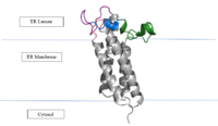
[[Image:VKORmembrane.PNG|200px|right|thumb|'''Figure 2: Vitamin K Epoxide Reductase in the Endoplasmic Membrane''']] | [[Image:VKORmembrane.PNG|200px|right|thumb|'''Figure 2: Vitamin K Epoxide Reductase in the Endoplasmic Membrane''']] | ||
| - | In the liver, the VKOR enzyme is set in the endoplasmic reticulum membrane (Fig.2). The transmembrane helices are located in the Endoplasmic Reticulum Luminal Region, which is the region between the ER Lumen and the [https://en.wikipedia.org/wiki/Cytosol Cytosol]. The cap region is partially oriented in the ER Lumen. The active site remains within the [https://en.wikipedia.org/wiki/Endoplasmic_reticulum Endoplasmic Reticulum Membrane].The Anchor is partially within the ER lumen, and partially embedded in the ER membrane. The anchor is what attaches the cap domain and stabilizes it, which allows the cap domain to cover the active site. | + | In the liver, the VKOR enzyme is set in the endoplasmic reticulum membrane (Fig.2). The transmembrane helices are located in the Endoplasmic Reticulum Luminal Region, which is the region between the ER Lumen and the [https://en.wikipedia.org/wiki/Cytosol Cytosol]. The cap region is partially oriented in the ER Lumen. The active site remains within the [https://en.wikipedia.org/wiki/Endoplasmic_reticulum Endoplasmic Reticulum Membrane].The Anchor is partially within the ER lumen, and partially embedded in the ER membrane. The anchor is what attaches the cap domain and stabilizes it, which allows the cap domain to cover the active site.<ref name=”Liu”>PMID:33154105</ref> |
| - | The VKOR enzyme is made up of four transmembrane helices: <scene name='90/904321/Tm1/1'>TM1</scene>, <scene name='90/904321/Tm2/2'>TM2</scene>, <scene name='90/904321/Tm3/2'>TM3</scene>, and <scene name='90/904321/Tm4/2'>TM4</scene> .(Grey/Orange) Each of these helices come together to form a central ligand binding pocket. This central pocket is the active site where conserved Cysteines: C132 and C135 are located. In the cap domain are important regions that are significant for Vitamin K binding, and the overall function of Vitamin K Epoxide Reductase, including the Anchor(Green), Cap Sequence (Blue), Beta Hairpin (Purple), and 3-4 Loop (Pink). | + | The VKOR enzyme is made up of four transmembrane helices: <scene name='90/904321/Tm1/1'>TM1</scene>, <scene name='90/904321/Tm2/2'>TM2</scene>, <scene name='90/904321/Tm3/2'>TM3</scene>, and <scene name='90/904321/Tm4/2'>TM4</scene> .(Grey/Orange) Each of these helices come together to form a central ligand binding pocket. This central pocket is the active site where conserved Cysteines: C132 and C135 are located. In the cap domain are important regions that are significant for Vitamin K binding, and the overall function of Vitamin K Epoxide Reductase, including the Anchor(Green), Cap Sequence (Blue), Beta Hairpin (Purple), and 3-4 Loop (Pink). <ref name=”Liu”>PMID:33154105</ref> |
| - | The <scene name='90/904321/Anchor/3'>Anchor</scene> attaches to the cap domain of the Vitamin K Epoxide Reductase Enzyme and is partially embedded in the Endoplasmic Reticulum Membrane. This both stabilizes the enzyme in the membrane, and stabilizes the cap domain over the active site. | + | The <scene name='90/904321/Anchor/3'>Anchor</scene> attaches to the cap domain of the Vitamin K Epoxide Reductase Enzyme and is partially embedded in the Endoplasmic Reticulum Membrane. This both stabilizes the enzyme in the membrane, and stabilizes the cap domain over the active site. <ref name=”Liu”>PMID:33154105</ref> |
| - | The <scene name='90/904321/Cap_sequence/1'>Cap Sequence</scene> is two parts: The cap helix and the cap loop. When the enzyme is reducing Vitamin K Epoxide or being inhibited by Vitamin K Antagonists, this cap region swings downward over the active site. The cap region is directly attached to the anchor. | + | The <scene name='90/904321/Cap_sequence/1'>Cap Sequence</scene> is two parts: The cap helix and the cap loop. When the enzyme is reducing Vitamin K Epoxide or being inhibited by Vitamin K Antagonists, this cap region swings downward over the active site. The cap region is directly attached to the anchor. <ref name=”Liu”>PMID:33154105</ref> |
''' | ''' | ||
| - | The <scene name='90/904321/Beta_hairpin/1'>Beta Hairpin</scene> is only seen in the closed conformation of Vitamin K Epoxide Reductase. When in the open conformation the beta hairpin is referred to as the luminal helix (yellow). The Beta hairpin is significant due to the fact that it contains the other two conserved cysteines necessary for the function of Vitamin K Epoxide Reductase: Cysteine43 and Cysteine51. The beta hairpin/luminal helix is directly connected to the cap region. | + | The <scene name='90/904321/Beta_hairpin/1'>Beta Hairpin</scene> is only seen in the closed conformation of Vitamin K Epoxide Reductase. When in the open conformation the beta hairpin is referred to as the luminal helix (yellow). The Beta hairpin is significant due to the fact that it contains the other two conserved cysteines necessary for the function of Vitamin K Epoxide Reductase: Cysteine43 and Cysteine51. The beta hairpin/luminal helix is directly connected to the cap region. <ref name=”Liu”>PMID:33154105</ref> |
| - | The <scene name='90/904321/3-4_loop/2'>Loop 3-4</scene> is the sequence of residues between Transmembrane Helix 3 and Transmembrane Helix 4. In the open conformation the loop does not have significant interactions with the rest of the cap domain, however in the closed conformation Loop 3-4 has many hydrogen reactions with the Cap Loop. This allows for the stabilization when VKOR is closed. | + | The <scene name='90/904321/3-4_loop/2'>Loop 3-4</scene> is the sequence of residues between Transmembrane Helix 3 and Transmembrane Helix 4. In the open conformation the loop does not have significant interactions with the rest of the cap domain, however in the closed conformation Loop 3-4 has many hydrogen reactions with the Cap Loop. This allows for the stabilization when VKOR is closed. <ref name=”Liu”>PMID:33154105</ref> |
== Vitamin K Epoxide == | == Vitamin K Epoxide == | ||
| Line 55: | Line 55: | ||
==Catalytic Cycle== | ==Catalytic Cycle== | ||
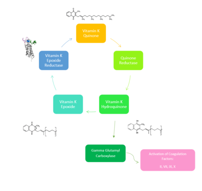
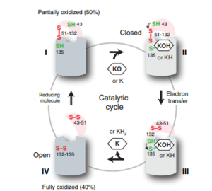
| - | [[Image:VKORcycle.PNG|300px|right|thumb|'''Figure 4: The catalytic cycle of Vitamin K Epoxide Reductase''' <ref name= | + | [[Image:VKORcycle.PNG|300px|right|thumb|'''Figure 4: The catalytic cycle of Vitamin K Epoxide Reductase''' <ref name=”Liu”>PMID:33154105</ref> ]] |
| Line 65: | Line 65: | ||
===Step I === | ===Step I === | ||
| - | <scene name='90/904321/I/1'>Step I</scene> of reforming Vitamin K Epoxide (Fig. 3) through the enzyme Vitamin K Reductase (VKOR) begins in a partially oxidized open conformation. In this state, catalytic cysteines 51 and 132 form a disulfide bond. Cysteines 43 and 135 are considered "free" because they are not bound to anything in this state. The <scene name='90/904321/I/2'>central binding pocket</scene> (highlighted in hot pink) is also empty because Vitamin K Epoxide has not bound yet. In order to get to the next step, Vitamin K epoxide will enter through the isoprenyl-chain tunnel.<ref name= | + | <scene name='90/904321/I/1'>Step I</scene> of reforming Vitamin K Epoxide (Fig. 3) through the enzyme Vitamin K Reductase (VKOR) begins in a partially oxidized open conformation. In this state, catalytic cysteines 51 and 132 form a disulfide bond. Cysteines 43 and 135 are considered "free" because they are not bound to anything in this state. The <scene name='90/904321/I/2'>central binding pocket</scene> (highlighted in hot pink) is also empty because Vitamin K Epoxide has not bound yet. In order to get to the next step, Vitamin K epoxide will enter through the isoprenyl-chain tunnel.<ref name=”Liu”>PMID:33154105</ref> |
===Step II=== | ===Step II=== | ||
| Line 74: | Line 74: | ||
===Step IV=== | ===Step IV=== | ||
| - | <scene name='90/904321/Iv/1'>Step IV</scene> is the last step of this cycle. Vitamin K Quinone will exit the central binding pocket and the open conformation will form. VKOR is in its fully oxidized state after donating its electrons to Vitamin K Epoxide. This is when the luminal helix will be visible. The cycle then repeats at Step I to keep reducing Vitamin K Epoxide to Vitamin K Quinone. | + | <scene name='90/904321/Iv/1'>Step IV</scene> is the last step of this cycle. Vitamin K Quinone will exit the central binding pocket and the open conformation will form. VKOR is in its fully oxidized state after donating its electrons to Vitamin K Epoxide. This is when the luminal helix will be visible. The cycle then repeats at Step I to keep reducing Vitamin K Epoxide to Vitamin K Quinone. <ref name=”Liu”>PMID:33154105</ref> |
Revision as of 14:10, 19 April 2022
Vitamin K Epoxide Reductase
| |||||||||||
References
- ↑ Shen G, Cui W, Cao Q, Gao M, Liu H, Su G, Gross ML, Li W. The catalytic mechanism of vitamin K epoxide reduction in a cellular environment. J Biol Chem. 2021 Jan-Jun;296:100145. doi: 10.1074/jbc.RA120.015401. Epub 2020, Dec 10. PMID:33273012 doi:http://dx.doi.org/10.1074/jbc.RA120.015401
- ↑ Liu S, Li S, Shen G, Sukumar N, Krezel AM, Li W. Structural basis of antagonizing the vitamin K catalytic cycle for anticoagulation. Science. 2020 Nov 5. pii: science.abc5667. doi: 10.1126/science.abc5667. PMID:33154105 doi:http://dx.doi.org/10.1126/science.abc5667
- ↑ Liu S, Li S, Shen G, Sukumar N, Krezel AM, Li W. Structural basis of antagonizing the vitamin K catalytic cycle for anticoagulation. Science. 2020 Nov 5. pii: science.abc5667. doi: 10.1126/science.abc5667. PMID:33154105 doi:http://dx.doi.org/10.1126/science.abc5667
- ↑ Liu S, Li S, Shen G, Sukumar N, Krezel AM, Li W. Structural basis of antagonizing the vitamin K catalytic cycle for anticoagulation. Science. 2020 Nov 5. pii: science.abc5667. doi: 10.1126/science.abc5667. PMID:33154105 doi:http://dx.doi.org/10.1126/science.abc5667
- ↑ Liu S, Li S, Shen G, Sukumar N, Krezel AM, Li W. Structural basis of antagonizing the vitamin K catalytic cycle for anticoagulation. Science. 2020 Nov 5. pii: science.abc5667. doi: 10.1126/science.abc5667. PMID:33154105 doi:http://dx.doi.org/10.1126/science.abc5667
- ↑ Liu S, Li S, Shen G, Sukumar N, Krezel AM, Li W. Structural basis of antagonizing the vitamin K catalytic cycle for anticoagulation. Science. 2020 Nov 5. pii: science.abc5667. doi: 10.1126/science.abc5667. PMID:33154105 doi:http://dx.doi.org/10.1126/science.abc5667
- ↑ Liu S, Li S, Shen G, Sukumar N, Krezel AM, Li W. Structural basis of antagonizing the vitamin K catalytic cycle for anticoagulation. Science. 2020 Nov 5. pii: science.abc5667. doi: 10.1126/science.abc5667. PMID:33154105 doi:http://dx.doi.org/10.1126/science.abc5667
- ↑ Liu S, Li S, Shen G, Sukumar N, Krezel AM, Li W. Structural basis of antagonizing the vitamin K catalytic cycle for anticoagulation. Science. 2020 Nov 5. pii: science.abc5667. doi: 10.1126/science.abc5667. PMID:33154105 doi:http://dx.doi.org/10.1126/science.abc5667
- ↑ Liu S, Li S, Shen G, Sukumar N, Krezel AM, Li W. Structural basis of antagonizing the vitamin K catalytic cycle for anticoagulation. Science. 2020 Nov 5. pii: science.abc5667. doi: 10.1126/science.abc5667. PMID:33154105 doi:http://dx.doi.org/10.1126/science.abc5667
- ↑ Liu S, Li S, Shen G, Sukumar N, Krezel AM, Li W. Structural basis of antagonizing the vitamin K catalytic cycle for anticoagulation. Science. 2020 Nov 5. pii: science.abc5667. doi: 10.1126/science.abc5667. PMID:33154105 doi:http://dx.doi.org/10.1126/science.abc5667
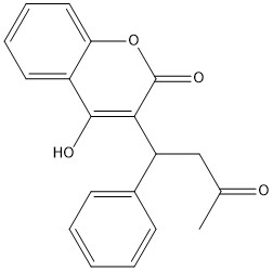
- ↑ PMID:23034830<ref></ref>Warfarin is the most common medication for this treatment, acting as a blood thinner. Warfarin binding to VKOR prevents the triggering of coagulation factors that form blood clots.
(DON’T INCLUDE)
[1] Goodstadt, L., & Ponting, C. P. (2004). Vitamin K epoxide reductase: homology, active site and catalytic mechanism. Trends in biochemical sciences, 29(6), 289–292. https://doi.org/10.1016/j.tibs.2004.04.004
Rishavy, M.A., Usubalieva, A., Hallgren, K.W., & Berkner, K.L. (2011). Novel insidht into the mechanism of the vitamin K oxidoreductas (VKOR): Electron relay through Cy43 and Cys51 reduces VKOR to allow vitamin K reduction and facilitation of vitamin K-dependent protein caroxylation. Journal of Biological Chemistry, 286(9), 7267-7278. https://doi.org/10.1074/jbc.M110.172213
[2] Liu, S., Li, S., Shen, G., Sukumar, N., Krezel, A. M., & Li, W. (2021). Structural basis of antagonizing the vitamin K catalytic cycle for anticoagulation. Science (New York, N.Y.), 371(6524), eabc5667. https://doi.org/10.1126/science.abc5667
[3] Shen, G., Cui, W., Cao, Q., Gao, M., Liu, H., Su, G., Gross, M. L., & Li, W. (2021). The catalytic mechanism of vitamin K epoxide reduction in a cellular environment. The Journal of biological chemistry, 296, 100145. https://doi.org/10.1074/jbc.RA120.015401
Silverman, R.B. (1981). Chemical model studies for the mechanism of vitamin K epoxide reductase. The Journal of American Chemistry Society, 103(19), 5939-5941. [4] [5] [6] [7] [8]
