This old version of Proteopedia is provided for student assignments while the new version is undergoing repairs. Content and edits done in this old version of Proteopedia after March 1, 2026 will eventually be lost when it is retired in about June of 2026.
Apply for new accounts at the new Proteopedia. Your logins will work in both the old and new versions.
2xoa
From Proteopedia
(Difference between revisions)
| Line 3: | Line 3: | ||
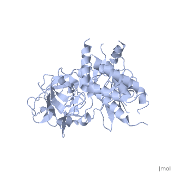
<StructureSection load='2xoa' size='340' side='right'caption='[[2xoa]], [[Resolution|resolution]] 2.50Å' scene=''> | <StructureSection load='2xoa' size='340' side='right'caption='[[2xoa]], [[Resolution|resolution]] 2.50Å' scene=''> | ||
== Structural highlights == | == Structural highlights == | ||
| - | <table><tr><td colspan='2'>[[2xoa]] is a 1 chain structure with sequence from [ | + | <table><tr><td colspan='2'>[[2xoa]] is a 1 chain structure with sequence from [https://en.wikipedia.org/wiki/European_rabbit European rabbit]. Full crystallographic information is available from [http://oca.weizmann.ac.il/oca-bin/ocashort?id=2XOA OCA]. For a <b>guided tour on the structure components</b> use [https://proteopedia.org/fgij/fg.htm?mol=2XOA FirstGlance]. <br> |
| - | </td></tr><tr id='related'><td class="sblockLbl"><b>[[Related_structure|Related:]]</b></td><td class="sblockDat">[[2bcx|2bcx]]</td></tr> | + | </td></tr><tr id='related'><td class="sblockLbl"><b>[[Related_structure|Related:]]</b></td><td class="sblockDat"><div style='overflow: auto; max-height: 3em;'>[[2bcx|2bcx]]</div></td></tr> |
| - | <tr id='resources'><td class="sblockLbl"><b>Resources:</b></td><td class="sblockDat"><span class='plainlinks'>[ | + | <tr id='resources'><td class="sblockLbl"><b>Resources:</b></td><td class="sblockDat"><span class='plainlinks'>[https://proteopedia.org/fgij/fg.htm?mol=2xoa FirstGlance], [http://oca.weizmann.ac.il/oca-bin/ocaids?id=2xoa OCA], [https://pdbe.org/2xoa PDBe], [https://www.rcsb.org/pdb/explore.do?structureId=2xoa RCSB], [https://www.ebi.ac.uk/pdbsum/2xoa PDBsum], [https://prosat.h-its.org/prosat/prosatexe?pdbcode=2xoa ProSAT]</span></td></tr> |
</table> | </table> | ||
== Function == | == Function == | ||
| - | [[ | + | [[https://www.uniprot.org/uniprot/RYR1_RABIT RYR1_RABIT]] Calcium channel that mediates the release of Ca(2+) from the sarcoplasmic reticulum into the cytoplasm and thereby plays a key role in triggering muscle contraction following depolarization of T-tubules. Repeated very high-level exercise increases the open probability of the channel and leads to Ca(2+) leaking into the cytoplasm. Can also mediate the release of Ca(2+) from intracellular stores in neurons, and may thereby promote prolonged Ca(2+) signaling in the brain. Required for normal embryonic development of muscle fibers and skeletal muscle. Required for normal heart morphogenesis, skin development and ossification during embryogenesis (By similarity).<ref>PMID:10388749</ref> <ref>PMID:22036948</ref> |
<div style="background-color:#fffaf0;"> | <div style="background-color:#fffaf0;"> | ||
== Publication Abstract from PubMed == | == Publication Abstract from PubMed == | ||
Revision as of 12:14, 27 April 2022
Crystal Structure of the N-terminal three domains of the skeletal muscle Ryanodine Receptor (RyR1)
| |||||||||||