We apologize for Proteopedia being slow to respond. For the past two years, a new implementation of Proteopedia has been being built. Soon, it will replace this 18-year old system. All existing content will be moved to the new system at a date that will be announced here.
Sandbox reserved 1752
From Proteopedia
(Difference between revisions)
| Line 18: | Line 18: | ||
<scene name='92/925553/Atp_analog/6'>ATP analog</scene> | <scene name='92/925553/Atp_analog/6'>ATP analog</scene> | ||
Stop here | Stop here | ||
| + | ==Cystic fibrosis transmembrane conductance regulator (CFTR)== | ||
| + | <StructureSection load='5UAK' size='340' side='right' caption='Cystic Fibrosis Transmembrane Conductance regulator' scene=''> | ||
| + | The '''CFTR''' is a chloride channel, and is regulated by PKA phosphorylation, cAMP levels, and ATP/ADP ratios. Mutations in the CFTR cause the disease cystic fibrosis. | ||
| + | |||
| + | CFTR is a mostly <scene name='78/785332/Secondary_structure/1'>alpha helical</scene> protein. The membrane spanning segments can be clearly seen with coloring by <scene name='78/785332/Hydrophobicity/1'>hydrophobicity</scene>, which shows hydrophobic residues in gray and hydrophilic residues in purple. | ||
| + | |||
| + | The extracellular end of the channel has several <scene name='78/785332/Ec_cl_selection/1'>positively charged</scene> residues that are important for recruiting chloride ions to the channel. A number of <scene name='78/785332/Plus_channel/1'>positively charged</scene> residues line the channel. In the unphosphorylated state (as this structure is), a <scene name='78/785332/Regulatory_domain/2'>regulatory domain</scene> blocks the activity of the channel (the connecting segments are not visible in the structure). It contains several negatively charged residues; when the protein is phosphorylated, this segment is repelled, causing a structural change. <ref>PMID:28340353</ref> | ||
| + | |||
| + | CFTR contains two <scene name='78/785332/Nbd/2'>nucleotide binding domains</scene> (NBD's), which both contain <scene name='78/785332/Walker_motifs/2'>Walker motifs</scene>, flexible loops that bind phosphate groups tightly and are highly conserved among ATP-binding proteins. | ||
| + | |||
| + | ==Mutations in Cystic Fibrosis== | ||
| + | |||
| + | Cystic fibrosis is characterized by decreased chloride transport, which causes mucus to be thicker and stickier. This leads to a variety of problems, including decreased lung capacity, decreased pancreatic enzyme release into the small intestine, increased rates of lung infections, and infertility.<ref>https://ghr.nlm.nih.gov/condition/cystic-fibrosis</ref> There are a wide assortment of mutations that cause cystic fibrosis, with differing symptom severity. The deletion of <scene name='78/785332/F508/1'>F508</scene> causes the protein to not be properly synthesized, and no expression is seen on the cell surface. Other mutations are found in the NBD's; some of these mutations such as S1255P alter the responsiveness to MgATP, while others such as G551S, G1244E, and G1239D decrease the frequency of channel opening. | ||
| + | |||
| + | Some of the mutations that lead to cystic fibrosis are due to folding errors. There are <scene name='78/785332/Numbered_bundles/2'>12 transmembrane sequences</scene> in CFTR; they are not sequential in their packing. The presence of <scene name='78/785332/Numbered_bundles_pos_res/1'>hydrophilic, positively charged amino acids</scene> in these transmembrane sequences (shown in red) lead to a folding problem: how do you stabilize them until they can be protected by hydrophobic residues and are no longer exposed to the hydrophobic membrane? | ||
| + | |||
| + | ==3D Printed Physical Model of the CFTR protein== | ||
| + | |||
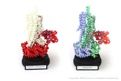
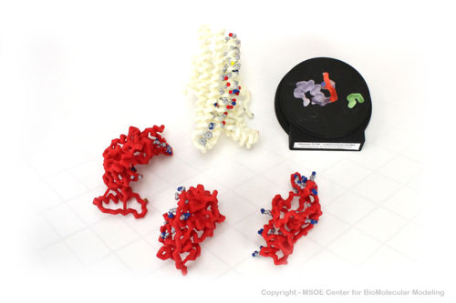
| + | Shown below are 3D printed physical models of the Cystic Fibrosis Transmembrane Conductance Regulator (CFTR) protein. The backbone model on the left is colored by region, with the transmembrane domain white and the atp-binding and regulatory domains colored red. The backbone model on the right is colored by regional repeat, with the first repeat blue, the second repeat green and the regulatory domain colored red. The models have been designed with embedded magnets to disassemble into the key regions of the structure. | ||
| + | |||
| + | [[Image:cftr1_centerForBioMolecularModeling.jpg|500px]] | ||
| + | [[Image:cftr2_centerForBioMolecularModeling.jpg|500px]] | ||
| + | |||
| + | ====The MSOE Center for BioMolecular Modeling==== | ||
| + | |||
| + | [[Image:CbmUniversityLogo.jpg | left | 150px]] | ||
| + | |||
| + | The [http://cbm.msoe.edu MSOE Center for BioMolecular Modeling] uses 3D printing technology to create physical models of protein and molecular structures, making the invisible molecular world more tangible and comprehensible. To view more protein structure models, visit our [http://cbm.msoe.edu/educationalmedia/modelgallery/ Model Gallery]. | ||
| + | |||
| + | |||
| + | |||
| + | </StructureSection> | ||
| + | |||
| + | |||
== References == | == References == | ||
<references /> | <references /> | ||
Current revision
| |||||||||||
References
- ↑ 1.0 1.1 1.2 1.3 1.4 Lee JY, Yang W. UvrD helicase unwinds DNA one base pair at a time by a two-part power stroke. Cell. 2006 Dec 29;127(7):1349-60. PMID:17190599 doi:http://dx.doi.org/10.1016/j.cell.2006.10.049
- ↑ Voet, D., Voet, J., & Pratt, C. (2015). Fundamentals of Biochemistry: Life at the Molecular Level (4th ed.). Wiley
- ↑ Liu F, Zhang Z, Csanady L, Gadsby DC, Chen J. Molecular Structure of the Human CFTR Ion Channel. Cell. 2017 Mar 23;169(1):85-95.e8. doi: 10.1016/j.cell.2017.02.024. PMID:28340353 doi:http://dx.doi.org/10.1016/j.cell.2017.02.024
- ↑ https://ghr.nlm.nih.gov/condition/cystic-fibrosis


