Sandbox Reserved 1781
From Proteopedia
(Difference between revisions)
| Line 11: | Line 11: | ||
== Structure == | == Structure == | ||
===Overview=== | ===Overview=== | ||
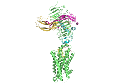
| - | The thyrotropin receptor has an extracellular domain (ECD) that is composed of a leucine rich repeat domain (LRRD) as well as the hinge region. This hinge region links the ECD to the seven transmembrane helices, which span from the extracellular domain to the intracellular domain <ref name= "Keinau et al.">PMID:228484426</ref>. When thyrotropin or an autoantibody binds, it causes a conformational change in the receptor through the transmembrane helices. This causes the thyrotropin receptor to interact differently with its respective G-protein when in the active and inactive states. | + | The thyrotropin receptor has an extracellular domain (ECD) that is composed of a <scene name='95/952709/Overview_of_the_ecd/1'>leucine rich repeat domain (LRRD)</scene> as well as the hinge region. This hinge region links the ECD to the seven transmembrane helices, which span from the extracellular domain to the intracellular domain <ref name= "Keinau et al.">PMID:228484426</ref>. When thyrotropin or an autoantibody binds, it causes a conformational change in the receptor through the transmembrane helices. This causes the thyrotropin receptor to interact differently with its respective G-protein when in the active and inactive states. |
===7 Transmembrane Helices=== | ===7 Transmembrane Helices=== | ||
The thyrotropin receptor is anchored to the membrane through seven transmembrane helices which is characteristic of | The thyrotropin receptor is anchored to the membrane through seven transmembrane helices which is characteristic of | ||
Revision as of 20:06, 24 March 2023
| This Sandbox is Reserved from February 27 through August 31, 2023 for use in the course CH462 Biochemistry II taught by R. Jeremy Johnson at the Butler University, Indianapolis, USA. This reservation includes Sandbox Reserved 1765 through Sandbox Reserved 1795. |
To get started:
More help: Help:Editing |
Your Heading Here (maybe something like 'Structure')
| |||||||||||
References
- ↑ Hanson, R. M., Prilusky, J., Renjian, Z., Nakane, T. and Sussman, J. L. (2013), JSmol and the Next-Generation Web-Based Representation of 3D Molecular Structure as Applied to Proteopedia. Isr. J. Chem., 53:207-216. doi:http://dx.doi.org/10.1002/ijch.201300024
- ↑ Herraez A. Biomolecules in the computer: Jmol to the rescue. Biochem Mol Biol Educ. 2006 Jul;34(4):255-61. doi: 10.1002/bmb.2006.494034042644. PMID:21638687 doi:10.1002/bmb.2006.494034042644
- ↑ Yen PM. Physiological and molecular basis of thyroid hormone action. Physiol Rev. 2001 Jul;81(3):1097-142. doi: 10.1152/physrev.2001.81.3.1097. PMID: 11427693.
- ↑ Duan J, Xu P, Luan X, Ji Y, He X, Song N, Yuan Q, Jin Y, Cheng X, Jiang H, Zheng J, Zhang S, Jiang Y, Xu HE. Hormone- and antibody-mediated activation of the thyrotropin receptor. Nature. 2022 Aug 8. pii: 10.1038/s41586-022-05173-3. doi:, 10.1038/s41586-022-05173-3. PMID:35940204 doi:http://dx.doi.org/10.1038/s41586-022-05173-3
- ↑ Kohn LD, Shimura H, Shimura Y, Hidaka A, Giuliani C, Napolitano G, Ohmori M, Laglia G, Saji M. The thyrotropin receptor. Vitam Horm. 1995;50:287-384. doi: 10.1016/s0083-6729(08)60658-5. PMID: 7709602.
- ↑ . PMID:228484426

