BASIL2023GVP76586
From Proteopedia
(Difference between revisions)
| Line 29: | Line 29: | ||
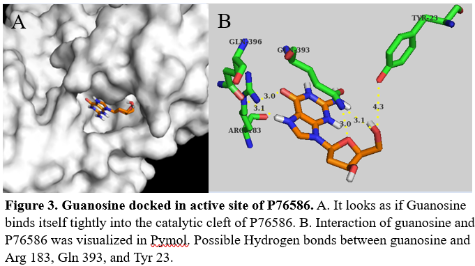
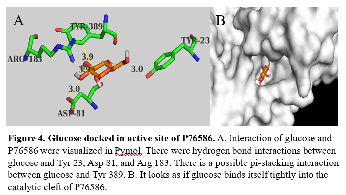
<scene name='95/957646/Globular_structure/1'>P76586 is a globular protein</scene> with 397 amino acids. It's <scene name='95/957646/Secondary_structure/1'>secondary structure</scene> is made up of mostly alpha helices, but also has beta sheets and random coil. Through docking we were able to identify possible amino acids involved in the <scene name='95/957646/Active_site/1'>active site</scene> of P76586. Potential amino acids in the active site are Tyr23, Asp81, Arg183, Gln393, and Tyr389. | <scene name='95/957646/Globular_structure/1'>P76586 is a globular protein</scene> with 397 amino acids. It's <scene name='95/957646/Secondary_structure/1'>secondary structure</scene> is made up of mostly alpha helices, but also has beta sheets and random coil. Through docking we were able to identify possible amino acids involved in the <scene name='95/957646/Active_site/1'>active site</scene> of P76586. Potential amino acids in the active site are Tyr23, Asp81, Arg183, Gln393, and Tyr389. | ||
== Results == | == Results == | ||
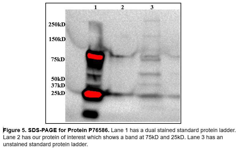
| - | Our protein of interest has a weight of ≈44.53kD. When analyzing SDS PAGE (figure 5) we were slightly concerned we weren't working with our protein of interest. We didn't get a great image out of SDS, if there were more time we would run again with more protein in the well so that we could see it better. We also made the mistake of not including our induction samples and our fractions from protein purification using | + | Our protein of interest has a weight of ≈44.53kD. When analyzing SDS PAGE (figure 5) we were slightly concerned we weren't working with our protein of interest. We didn't get a great image out of SDS, if there were more time we would run again with more protein in the well so that we could see it better. We also made the mistake of not including our induction samples and our fractions from protein purification using Nickle affinity chromatography. The band at 75kD and 25kD we believe are there from the imaging and is not from our actual protein sample. |
[[Image:SDS.png]] | [[Image:SDS.png]] | ||
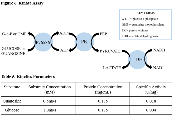
| - | We ran two coupled kinase assays, one with glucose and one with guanosine. After calculating the specific activity of our protein with glucose and guanosine we determined there was not enough activity for glucose or guanosine to be the substrate (table 5). | + | We ran two coupled kinase assays, one with glucose and one with guanosine. After calculating the specific activity of our protein with glucose and guanosine we determined there was not enough activity for glucose or guanosine to be the substrate (table 5). We didn't have to modify the kinase assay protocol for Abl Kinase other than changing the substrate and ensuring we were using a high enough substrate concentration that was greater than the protein concentration. |
[[Image:Kinase_assay.png]] | [[Image:Kinase_assay.png]] | ||
Revision as of 15:42, 26 April 2023
Investigating the Function of Protein P76586
| |||||||||||
References
- ↑ Hanson, R. M., Prilusky, J., Renjian, Z., Nakane, T. and Sussman, J. L. (2013), JSmol and the Next-Generation Web-Based Representation of 3D Molecular Structure as Applied to Proteopedia. Isr. J. Chem., 53:207-216. doi:http://dx.doi.org/10.1002/ijch.201300024
- ↑ Herraez A. Biomolecules in the computer: Jmol to the rescue. Biochem Mol Biol Educ. 2006 Jul;34(4):255-61. doi: 10.1002/bmb.2006.494034042644. PMID:21638687 doi:10.1002/bmb.2006.494034042644
