Function
3-phosphoinositide-dependent protein kinase 1 (PDK1) activates many other kinases. PDK1 main effector is protein kinase B (Akt). . PDK1 is important in signaling pathways activated by growth factors and hormones. It interacts with membrane phospholipids. [1]
PDK1 is an enzyme that plays a central role in signal transduction pathways involved in cell growth, metabolism, and survival. It belongs to the family of AGC (protein kinase A, G, and C) kinases and is a key regulator of various cellular processes.
PDK1 is known for its ability to phosphorylate and activate a group of kinases called AGC kinases, which includes protein kinase B (PKB/Akt), protein kinase C (PKC), and serum and glucocorticoid-regulated kinase (SGK), among others. PDK1 phosphorylates a conserved activation loop residue (threonine or serine) of AGC kinases, which leads to their activation and subsequent downstream signaling events.
The activation of PDK1 is tightly regulated and relies on the presence of phosphatidylinositol-3,4,5-trisphosphate (PIP3) or phosphatidylinositol-3,4-bisphosphate (PIP2), which are generated by phosphoinositide 3-kinase (PI3K) signaling. PDK1 contains a pleckstrin homology (PH) domain that specifically binds to these phosphoinositides, allowing its recruitment to the plasma membrane, where it becomes active.
Once activated, PDK1 phosphorylates the AGC kinases on a conserved residue within their activation loop, initiating a signaling cascade that regulates multiple cellular processes. These processes include cell proliferation, growth, survival, metabolism, and cytoskeletal rearrangements.
The dysregulation of PDK1 and its downstream signaling pathways has been implicated in various diseases, including cancer, diabetes, and cardiovascular disorders. Consequently, PDK1 has emerged as a potential therapeutic target for the development of novel drugs aimed at modulating these signaling pathways.
In summary, 3-phosphoinositide-dependent protein kinase 1 (PDK1) is a crucial enzyme involved in the activation of AGC kinases, which regulate a wide range of cellular processes. Its activation is dependent on phosphoinositide signaling, and it serves as a central hub in various signaling pathways involved in cell growth, metabolism, and survival.
Disease
Inhibition of PDK1 leads to increase in α-secretase activity at the neuronal surface causing cellular prion protein and amyloid precursor protein to be cleaved into their nonpathogenic forms.
Structural highlights
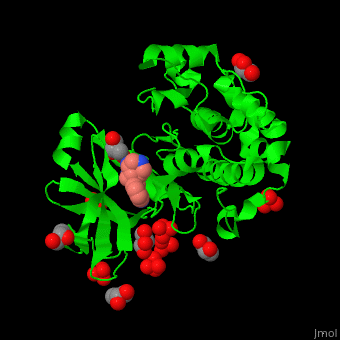
PDK1 structure contains 2 domains: kinase (residues 71-359 in human) and PH (residues 459-550) in human). The PH (Plecksin Homology) domain interacts with phospholipids. The kinase domain (kd) contains 3 binding sites. These are for substrate-binding, ATP-binding and allosteric activator docking (PIF - [PDK1-Interacting Fragment] -pocket). . Water molecules shown as red spheres.
3D Structures of PDK1
Pdk1 3D structures