Introduction
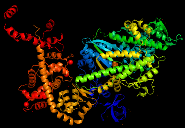
This is a figure of Myosin VI structure in gradient rainbow representation, where blue is N-terminal (5’) and red the C-terminal (3’).

Function
Predicted amino acid sequences and cDNA comparison of Myosin VI in different species shows strong evolutionary conservation, mainly in the head/motor domain and in the distal tail region. This is probably due to the unique and different functions of this protein. This scene represents the , where pink gradient shows the more conserved region, white is average, blue gradient is the variable region and grey or yellow is insufficient data.
Structure highlights
The crystal structure of the entry '2BKI' was determined by x-ray diffraction with the resolution of 2,90 Å and is composed by 3 chains: and two chains of .
The motor domain contains two inserts that are unique in the myosin superfamily:
Insert 1: .
Location: This insert belongs to the U50kDa subdomain and it is located near the nucleotide-binding pocket and the Switch I.
Function: This insert provides unique kinetic characteristics: it modulates nucleotide binding and switch 1 flexibility, therefore, it slows ADP release and ATP-induced dissociation of the motor from actin (at saturating ATP concentrations).
Mechanism: As also seen in all other myosins, the conformation of switch I relative to the U50kDa subdomain is not altered by the presence of insert 1. However, the small loop ( - in grey) that follows this insert, is repositioned, standing out in the nucleotide-binding pocket (decreasing nucleotide accessibility by steric impediment ) and strongly interacting with switch 1 by the residues: (in red). Also, (in green) of insert 1 interacts with switch 1. (highlighted in blue) is specifically important because its position selectively interfere with ATP binding, while having little or no effect on ADP binding. Mutation of leucine 310 to glycine removes all influence of insert-1 on ATP binding.
Insert 2:
Location: This insert is between the converter and the IQ motif
Function: Redirectionare the lever arm and contains a new CaM-binding motif.
Mechanism: The proximal part of insert 2 ( - in red) wraps around the converter, while the distal part ( - in green) forms a CaM-binding motif. The insert 2 and its associated CaM molecule (with 4Ca2+), make specific interactions with the converter, many involving a variable loop ( - in magenta). The result of interactions is that IQ helix emerges ~120° from the position that it emerges in all other myosins, redirecting the IQ helix and the CaM towards the minus end of the actin filament.
3D structure
All 3D structure, models and scenes were based on 2BKI - Myosin VI nucleotide-free (MDinsert2-IQ) crystal structure, published in https://doi.org/10.1038/nature03592 - Ménétrey, J., Bahloul, A., Wells, A. L., Yengo, C. M., Morris, C. A., Sweeney, H. L., & Houdusse, A. (2005). The structure of the myosin VI motor reveals the mechanism of directionality reversal. Nature, 435(7043), 779–785.
References


