Aquaporin-1
From Proteopedia
(Difference between revisions)
| Line 17: | Line 17: | ||
{{Clear}} | {{Clear}} | ||
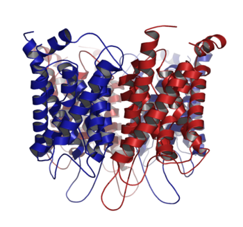
An AQP1 monomer unit contains six highly tilted transmembrane α-helices and two internal α-helices, oriented in the form of a barrel. The transmembrane α-helices thread through the membrane so as to expose both the N- and the C-terminus to the inner cytoplasm. The aqueous pathway within the monomer unit is characterized by a narrow curvilinear pore that is ~4.0Å in diameter, ~18Å in length, and bends ~25 degrees as it transverses the membrane.<ref name="structure" /> The curvilinear pore is a size selective pathway for water that excludes other larger and smaller molecules. The entrances to the aqueous pathway are mostly lined with polar and charged residues. Conversely, the interior of the aqueous pathway is mostly constituted by nonpolar residues. This feature allows for a relatively noninteracting pathway for the diffusion of water. | An AQP1 monomer unit contains six highly tilted transmembrane α-helices and two internal α-helices, oriented in the form of a barrel. The transmembrane α-helices thread through the membrane so as to expose both the N- and the C-terminus to the inner cytoplasm. The aqueous pathway within the monomer unit is characterized by a narrow curvilinear pore that is ~4.0Å in diameter, ~18Å in length, and bends ~25 degrees as it transverses the membrane.<ref name="structure" /> The curvilinear pore is a size selective pathway for water that excludes other larger and smaller molecules. The entrances to the aqueous pathway are mostly lined with polar and charged residues. Conversely, the interior of the aqueous pathway is mostly constituted by nonpolar residues. This feature allows for a relatively noninteracting pathway for the diffusion of water. | ||
| + | |||
| + | To visualize the channel, we can place pseudoatoms into it (using the [[PACUPP]] scripts). There is a bottleneck in the channel that acts as a filter. Cations like potassium or sodium ions, even though they are smaller than water, do not pass because together with a layer of hydration, they are larger than water. | ||
The N- and C-terminal halves of AQP1 are tandem repeats that contain a conserved <scene name='56/560863/Npa/3'>Asn-Pro-Ala (NPA)</scene> tripeptide sequence.<ref name="structure" /> Each terminal half is composed of three tilted transmembrane α-helices and a short α-helix adjacent to the conserved NPA motif. The two halves are connected through transmembrane 3 (TM3) and TM4 by a <scene name='56/560863/Interhelix_chain/1'>long interhelix chain</scene>. The location of the two NPA tripeptide sequences define the apex of the curvilinear pathway (N76 & N192), which is located about the center of the membrane. This orientation of helices allows for a sufficient amount of noncovalent interactions to induce stability. Interactions between helices are dominantly hydrophobic. However, hydrogen-bond interactions have also been suggested to increase stability. | The N- and C-terminal halves of AQP1 are tandem repeats that contain a conserved <scene name='56/560863/Npa/3'>Asn-Pro-Ala (NPA)</scene> tripeptide sequence.<ref name="structure" /> Each terminal half is composed of three tilted transmembrane α-helices and a short α-helix adjacent to the conserved NPA motif. The two halves are connected through transmembrane 3 (TM3) and TM4 by a <scene name='56/560863/Interhelix_chain/1'>long interhelix chain</scene>. The location of the two NPA tripeptide sequences define the apex of the curvilinear pathway (N76 & N192), which is located about the center of the membrane. This orientation of helices allows for a sufficient amount of noncovalent interactions to induce stability. Interactions between helices are dominantly hydrophobic. However, hydrogen-bond interactions have also been suggested to increase stability. | ||
Current revision
| |||||||||||
References
- ↑ Benga G. The first discovered water channel protein, later called aquaporin 1: molecular characteristics, functions and medical implications. Mol Aspects Med. 2012 Oct-Dec;33(5-6):518-34. doi: 10.1016/j.mam.2012.06.001., Epub 2012 Jun 15. PMID:22705445 doi:http://dx.doi.org/10.1016/j.mam.2012.06.001
- ↑ 2.0 2.1 Rutkovskiy A, Valen G, Vaage J. Cardiac aquaporins. Basic Res Cardiol. 2013 Nov;108(6):393. doi: 10.1007/s00395-013-0393-6. Epub 2013, Oct 25. PMID:24158693 doi:http://dx.doi.org/10.1007/s00395-013-0393-6
- ↑ Conner MT, Conner AC, Bland CE, Taylor LH, Brown JE, Parri HR, Bill RM. Rapid aquaporin translocation regulates cellular water flow: mechanism of hypotonicity-induced subcellular localization of aquaporin 1 water channel. J Biol Chem. 2012 Mar 30;287(14):11516-25. doi: 10.1074/jbc.M111.329219. Epub, 2012 Feb 9. PMID:22334691 doi:http://dx.doi.org/10.1074/jbc.M111.329219
- ↑ 4.0 4.1 4.2 4.3 Ren G, Reddy VS, Cheng A, Melnyk P, Mitra AK. Visualization of a water-selective pore by electron crystallography in vitreous ice. Proc Natl Acad Sci U S A. 2001 Feb 13;98(4):1398-403. Epub 2001 Jan 30. PMID:11171962 doi:10.1073/pnas.041489198
- ↑ Zhang W, Zitron E, Homme M, Kihm L, Morath C, Scherer D, Hegge S, Thomas D, Schmitt CP, Zeier M, Katus H, Karle C, Schwenger V. Aquaporin-1 channel function is positively regulated by protein kinase C. J Biol Chem. 2007 Jul 20;282(29):20933-40. Epub 2007 May 23. PMID:17522053 doi:http://dx.doi.org/10.1074/jbc.M703858200
Proteopedia Page Contributors and Editors (what is this?)
Alexander Berchansky, Karsten Theis, Kristopher Kleski, Michal Harel

