This old version of Proteopedia is provided for student assignments while the new version is undergoing repairs. Content and edits done in this old version of Proteopedia after March 1, 2026 will eventually be lost when it is retired in about June of 2026.
Apply for new accounts at the new Proteopedia. Your logins will work in both the old and new versions.
2e9s
From Proteopedia
(Difference between revisions)
| Line 3: | Line 3: | ||
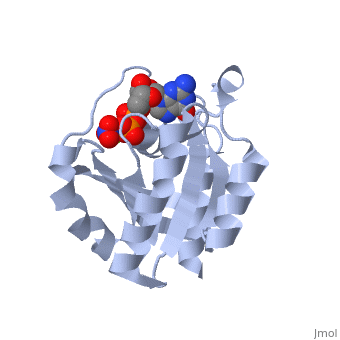
<StructureSection load='2e9s' size='340' side='right'caption='[[2e9s]], [[Resolution|resolution]] 1.78Å' scene=''> | <StructureSection load='2e9s' size='340' side='right'caption='[[2e9s]], [[Resolution|resolution]] 1.78Å' scene=''> | ||
== Structural highlights == | == Structural highlights == | ||
| - | <table><tr><td colspan='2'>[[2e9s]] is a 3 chain structure with sequence from [https://en.wikipedia.org/wiki/ | + | <table><tr><td colspan='2'>[[2e9s]] is a 3 chain structure with sequence from [https://en.wikipedia.org/wiki/Homo_sapiens Homo sapiens]. Full crystallographic information is available from [http://oca.weizmann.ac.il/oca-bin/ocashort?id=2E9S OCA]. For a <b>guided tour on the structure components</b> use [https://proteopedia.org/fgij/fg.htm?mol=2E9S FirstGlance]. <br> |
| - | </td></tr><tr id=' | + | </td></tr><tr id='method'><td class="sblockLbl"><b>[[Empirical_models|Method:]]</b></td><td class="sblockDat" id="methodDat">X-ray diffraction, [[Resolution|Resolution]] 1.78Å</td></tr> |
| - | <tr id=' | + | <tr id='ligand'><td class="sblockLbl"><b>[[Ligand|Ligands:]]</b></td><td class="sblockDat" id="ligandDat"><scene name='pdbligand=GDP:GUANOSINE-5-DIPHOSPHATE'>GDP</scene>, <scene name='pdbligand=MG:MAGNESIUM+ION'>MG</scene>, <scene name='pdbligand=NO3:NITRATE+ION'>NO3</scene></td></tr> |
| - | < | + | |
<tr id='resources'><td class="sblockLbl"><b>Resources:</b></td><td class="sblockDat"><span class='plainlinks'>[https://proteopedia.org/fgij/fg.htm?mol=2e9s FirstGlance], [http://oca.weizmann.ac.il/oca-bin/ocaids?id=2e9s OCA], [https://pdbe.org/2e9s PDBe], [https://www.rcsb.org/pdb/explore.do?structureId=2e9s RCSB], [https://www.ebi.ac.uk/pdbsum/2e9s PDBsum], [https://prosat.h-its.org/prosat/prosatexe?pdbcode=2e9s ProSAT]</span></td></tr> | <tr id='resources'><td class="sblockLbl"><b>Resources:</b></td><td class="sblockDat"><span class='plainlinks'>[https://proteopedia.org/fgij/fg.htm?mol=2e9s FirstGlance], [http://oca.weizmann.ac.il/oca-bin/ocaids?id=2e9s OCA], [https://pdbe.org/2e9s PDBe], [https://www.rcsb.org/pdb/explore.do?structureId=2e9s RCSB], [https://www.ebi.ac.uk/pdbsum/2e9s PDBsum], [https://prosat.h-its.org/prosat/prosatexe?pdbcode=2e9s ProSAT]</span></td></tr> | ||
</table> | </table> | ||
== Function == | == Function == | ||
| - | + | [https://www.uniprot.org/uniprot/RAB6B_HUMAN RAB6B_HUMAN] Seems to have a role in retrograde membrane traffic at the level of the Golgi complex. May function in retrograde transport in neuronal cells.<ref>PMID:17707369</ref> | |
== Evolutionary Conservation == | == Evolutionary Conservation == | ||
[[Image:Consurf_key_small.gif|200px|right]] | [[Image:Consurf_key_small.gif|200px|right]] | ||
| Line 21: | Line 20: | ||
</jmol>, as determined by [http://consurfdb.tau.ac.il/ ConSurfDB]. You may read the [[Conservation%2C_Evolutionary|explanation]] of the method and the full data available from [http://bental.tau.ac.il/new_ConSurfDB/main_output.php?pdb_ID=2e9s ConSurf]. | </jmol>, as determined by [http://consurfdb.tau.ac.il/ ConSurfDB]. You may read the [[Conservation%2C_Evolutionary|explanation]] of the method and the full data available from [http://bental.tau.ac.il/new_ConSurfDB/main_output.php?pdb_ID=2e9s ConSurf]. | ||
<div style="clear:both"></div> | <div style="clear:both"></div> | ||
| - | <div style="background-color:#fffaf0;"> | ||
| - | == Publication Abstract from PubMed == | ||
| - | The Rab small G-protein family plays important roles in eukaryotes as regulators of vesicle traffic. In Rab proteins, the hydrolysis of GTP to GDP is coupled with association with and dissociation from membranes. Conformational changes related to their different nucleotide states determine their effector specificity. The crystal structure of human neuronal Rab6B was solved in its 'inactive' (with bound MgGDP) and 'active' (MgGTPgammaS-bound) forms to 2.3 and 1.8 A, respectively. Both crystallized in space group P2(1)2(1)2(1), with similar unit-cell parameters, allowing the comparison of both structures without packing artifacts. Conformational changes between the inactive GDP and active GTP-like state are observed mainly in the switch I and switch II regions, confirming their role as a molecular switch. Compared with other Rab proteins, additional changes are observed in the Rab6 subfamily-specific RabSF3 region that might contribute to the specificity of Rab6 for its different effector proteins. | ||
| - | |||
| - | The structure of human neuronal Rab6B in the active and inactive form.,Garcia-Saez I, Tcherniuk S, Kozielski F Acta Crystallogr D Biol Crystallogr. 2006 Jul;62(Pt 7):725-33. Epub 2006, Jun 20. PMID:16790928<ref>PMID:16790928</ref> | ||
| - | |||
| - | From MEDLINE®/PubMed®, a database of the U.S. National Library of Medicine.<br> | ||
| - | </div> | ||
| - | <div class="pdbe-citations 2e9s" style="background-color:#fffaf0;"></div> | ||
==See Also== | ==See Also== | ||
| Line 37: | Line 27: | ||
__TOC__ | __TOC__ | ||
</StructureSection> | </StructureSection> | ||
| - | [[Category: | + | [[Category: Homo sapiens]] |
[[Category: Large Structures]] | [[Category: Large Structures]] | ||
| - | [[Category: Garcia-Saez | + | [[Category: Garcia-Saez I]] |
| - | [[Category: Kozielski | + | [[Category: Kozielski F]] |
| - | [[Category: Tcherniuk | + | [[Category: Tcherniuk S]] |
| - | [[Category: Vellieux | + | [[Category: Vellieux FM]] |
| - | + | ||
| - | + | ||
| - | + | ||
| - | + | ||
| - | + | ||
| - | + | ||
Current revision
human neuronal Rab6B in three intermediate forms
| |||||||||||