2xsc
From Proteopedia
(Difference between revisions)
| Line 4: | Line 4: | ||
== Structural highlights == | == Structural highlights == | ||
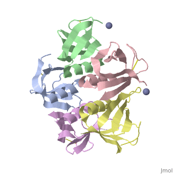
<table><tr><td colspan='2'>[[2xsc]] is a 5 chain structure with sequence from [https://en.wikipedia.org/wiki/Escherichia_coli Escherichia coli]. This structure supersedes the now removed PDB entry [http://oca.weizmann.ac.il/oca-bin/send-pdb?obs=1&id=1bov 1bov]. Full crystallographic information is available from [http://oca.weizmann.ac.il/oca-bin/ocashort?id=2XSC OCA]. For a <b>guided tour on the structure components</b> use [https://proteopedia.org/fgij/fg.htm?mol=2XSC FirstGlance]. <br> | <table><tr><td colspan='2'>[[2xsc]] is a 5 chain structure with sequence from [https://en.wikipedia.org/wiki/Escherichia_coli Escherichia coli]. This structure supersedes the now removed PDB entry [http://oca.weizmann.ac.il/oca-bin/send-pdb?obs=1&id=1bov 1bov]. Full crystallographic information is available from [http://oca.weizmann.ac.il/oca-bin/ocashort?id=2XSC OCA]. For a <b>guided tour on the structure components</b> use [https://proteopedia.org/fgij/fg.htm?mol=2XSC FirstGlance]. <br> | ||
| - | </td></tr><tr id=' | + | </td></tr><tr id='method'><td class="sblockLbl"><b>[[Empirical_models|Method:]]</b></td><td class="sblockDat" id="methodDat">X-ray diffraction, [[Resolution|Resolution]] 2.052Å</td></tr> |
| - | <tr id=' | + | <tr id='ligand'><td class="sblockLbl"><b>[[Ligand|Ligands:]]</b></td><td class="sblockDat" id="ligandDat"><scene name='pdbligand=ZN:ZINC+ION'>ZN</scene></td></tr> |
<tr id='resources'><td class="sblockLbl"><b>Resources:</b></td><td class="sblockDat"><span class='plainlinks'>[https://proteopedia.org/fgij/fg.htm?mol=2xsc FirstGlance], [http://oca.weizmann.ac.il/oca-bin/ocaids?id=2xsc OCA], [https://pdbe.org/2xsc PDBe], [https://www.rcsb.org/pdb/explore.do?structureId=2xsc RCSB], [https://www.ebi.ac.uk/pdbsum/2xsc PDBsum], [https://prosat.h-its.org/prosat/prosatexe?pdbcode=2xsc ProSAT]</span></td></tr> | <tr id='resources'><td class="sblockLbl"><b>Resources:</b></td><td class="sblockDat"><span class='plainlinks'>[https://proteopedia.org/fgij/fg.htm?mol=2xsc FirstGlance], [http://oca.weizmann.ac.il/oca-bin/ocaids?id=2xsc OCA], [https://pdbe.org/2xsc PDBe], [https://www.rcsb.org/pdb/explore.do?structureId=2xsc RCSB], [https://www.ebi.ac.uk/pdbsum/2xsc PDBsum], [https://prosat.h-its.org/prosat/prosatexe?pdbcode=2xsc ProSAT]</span></td></tr> | ||
</table> | </table> | ||
== Function == | == Function == | ||
| - | + | [https://www.uniprot.org/uniprot/STXB_BPH19 STXB_BPH19] The B subunit is responsible for the binding of the holotoxin to specific receptors on the target cell surface, such as globotriaosylceramide (Gb3) in human intestinal microvilli. | |
<div style="background-color:#fffaf0;"> | <div style="background-color:#fffaf0;"> | ||
== Publication Abstract from PubMed == | == Publication Abstract from PubMed == | ||
| Line 29: | Line 29: | ||
[[Category: Escherichia coli]] | [[Category: Escherichia coli]] | ||
[[Category: Large Structures]] | [[Category: Large Structures]] | ||
| - | [[Category: Boodhoo | + | [[Category: Boodhoo A]] |
| - | [[Category: Brunton | + | [[Category: Brunton JL]] |
| - | [[Category: Bunkoczi | + | [[Category: Bunkoczi G]] |
| - | [[Category: Oeffner | + | [[Category: Oeffner RD]] |
| - | [[Category: Read | + | [[Category: Read RJ]] |
| - | [[Category: Stein | + | [[Category: Stein PE]] |
| - | [[Category: Tyrrell | + | [[Category: Tyrrell GJ]] |
| - | + | ||
Current revision
Crystal structure of the cell-binding B oligomer of verotoxin-1 from E. coli
| |||||||||||