This old version of Proteopedia is provided for student assignments while the new version is undergoing repairs. Content and edits done in this old version of Proteopedia after March 1, 2026 will eventually be lost when it is retired in about June of 2026.
Apply for new accounts at the new Proteopedia. Your logins will work in both the old and new versions.
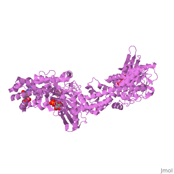
1cza
From Proteopedia
(Difference between revisions)
| Line 3: | Line 3: | ||
<StructureSection load='1cza' size='340' side='right'caption='[[1cza]], [[Resolution|resolution]] 1.90Å' scene=''> | <StructureSection load='1cza' size='340' side='right'caption='[[1cza]], [[Resolution|resolution]] 1.90Å' scene=''> | ||
== Structural highlights == | == Structural highlights == | ||
| - | <table><tr><td colspan='2'>[[1cza]] is a 1 chain structure with sequence from [https://en.wikipedia.org/wiki/ | + | <table><tr><td colspan='2'>[[1cza]] is a 1 chain structure with sequence from [https://en.wikipedia.org/wiki/Homo_sapiens Homo sapiens]. Full crystallographic information is available from [http://oca.weizmann.ac.il/oca-bin/ocashort?id=1CZA OCA]. For a <b>guided tour on the structure components</b> use [https://proteopedia.org/fgij/fg.htm?mol=1CZA FirstGlance]. <br> |
| - | </td></tr><tr id=' | + | </td></tr><tr id='method'><td class="sblockLbl"><b>[[Empirical_models|Method:]]</b></td><td class="sblockDat" id="methodDat">X-ray diffraction, [[Resolution|Resolution]] 1.9Å</td></tr> |
| - | <tr id=' | + | <tr id='ligand'><td class="sblockLbl"><b>[[Ligand|Ligands:]]</b></td><td class="sblockDat" id="ligandDat"><scene name='pdbligand=ADP:ADENOSINE-5-DIPHOSPHATE'>ADP</scene>, <scene name='pdbligand=G6P:ALPHA-D-GLUCOSE-6-PHOSPHATE'>G6P</scene>, <scene name='pdbligand=GLC:ALPHA-D-GLUCOSE'>GLC</scene></td></tr> |
| - | + | ||
<tr id='resources'><td class="sblockLbl"><b>Resources:</b></td><td class="sblockDat"><span class='plainlinks'>[https://proteopedia.org/fgij/fg.htm?mol=1cza FirstGlance], [http://oca.weizmann.ac.il/oca-bin/ocaids?id=1cza OCA], [https://pdbe.org/1cza PDBe], [https://www.rcsb.org/pdb/explore.do?structureId=1cza RCSB], [https://www.ebi.ac.uk/pdbsum/1cza PDBsum], [https://prosat.h-its.org/prosat/prosatexe?pdbcode=1cza ProSAT]</span></td></tr> | <tr id='resources'><td class="sblockLbl"><b>Resources:</b></td><td class="sblockDat"><span class='plainlinks'>[https://proteopedia.org/fgij/fg.htm?mol=1cza FirstGlance], [http://oca.weizmann.ac.il/oca-bin/ocaids?id=1cza OCA], [https://pdbe.org/1cza PDBe], [https://www.rcsb.org/pdb/explore.do?structureId=1cza RCSB], [https://www.ebi.ac.uk/pdbsum/1cza PDBsum], [https://prosat.h-its.org/prosat/prosatexe?pdbcode=1cza ProSAT]</span></td></tr> | ||
</table> | </table> | ||
== Disease == | == Disease == | ||
| - | + | [https://www.uniprot.org/uniprot/HXK1_HUMAN HXK1_HUMAN] Defects in HK1 are the cause of hexokinase deficiency (HK deficiency) [MIM:[https://omim.org/entry/235700 235700]. HK deficiency is a rare autosomal recessive disease with nonspherocytic hemolytic anemia as the predominant clinical feature. | |
| + | == Function == | ||
| + | [https://www.uniprot.org/uniprot/HXK1_HUMAN HXK1_HUMAN] | ||
== Evolutionary Conservation == | == Evolutionary Conservation == | ||
[[Image:Consurf_key_small.gif|200px|right]] | [[Image:Consurf_key_small.gif|200px|right]] | ||
| Line 21: | Line 22: | ||
</jmol>, as determined by [http://consurfdb.tau.ac.il/ ConSurfDB]. You may read the [[Conservation%2C_Evolutionary|explanation]] of the method and the full data available from [http://bental.tau.ac.il/new_ConSurfDB/main_output.php?pdb_ID=1cza ConSurf]. | </jmol>, as determined by [http://consurfdb.tau.ac.il/ ConSurfDB]. You may read the [[Conservation%2C_Evolutionary|explanation]] of the method and the full data available from [http://bental.tau.ac.il/new_ConSurfDB/main_output.php?pdb_ID=1cza ConSurf]. | ||
<div style="clear:both"></div> | <div style="clear:both"></div> | ||
| - | <div style="background-color:#fffaf0;"> | ||
| - | == Publication Abstract from PubMed == | ||
| - | Hexokinase I, the pacemaker of glycolysis in brain tissue, is composed of two structurally similar halves connected by an alpha-helix. The enzyme dimerizes at elevated protein concentrations in solution and in crystal structures; however, almost all published data reflect the properties of a hexokinase I monomer in solution. Crystal structures of mutant forms of recombinant human hexokinase I, presented here, reveal the enzyme monomer for the first time. The mutant hexokinases bind both glucose 6-phosphate and glucose with high affinity to their N and C-terminal halves, and ADP, also with high affinity, to a site near the N terminus of the polypeptide chain. Exposure of the monomer crystals to ADP in the complete absence of glucose 6-phosphate reveals a second binding site for adenine nucleotides at the putative active site (C-half), with conformational changes extending 15 A to the contact interface between the N and C-halves. The structures reveal distinct conformational states for the C-half and a rigid-body rotation of the N-half, as possible elements of a structure-based mechanism for allosteric regulation of catalysis. | ||
| - | |||
| - | Crystal structures of mutant monomeric hexokinase I reveal multiple ADP binding sites and conformational changes relevant to allosteric regulation.,Aleshin AE, Kirby C, Liu X, Bourenkov GP, Bartunik HD, Fromm HJ, Honzatko RB J Mol Biol. 2000 Mar 3;296(4):1001-15. PMID:10686099<ref>PMID:10686099</ref> | ||
| - | |||
| - | From MEDLINE®/PubMed®, a database of the U.S. National Library of Medicine.<br> | ||
| - | </div> | ||
| - | <div class="pdbe-citations 1cza" style="background-color:#fffaf0;"></div> | ||
==See Also== | ==See Also== | ||
*[[Hexokinase 3D structures|Hexokinase 3D structures]] | *[[Hexokinase 3D structures|Hexokinase 3D structures]] | ||
| - | == References == | ||
| - | <references/> | ||
__TOC__ | __TOC__ | ||
</StructureSection> | </StructureSection> | ||
| - | [[Category: | + | [[Category: Homo sapiens]] |
| - | + | ||
[[Category: Large Structures]] | [[Category: Large Structures]] | ||
| - | [[Category: Aleshin | + | [[Category: Aleshin AE]] |
| - | [[Category: Bartunik | + | [[Category: Bartunik HD]] |
| - | [[Category: Bourenkov | + | [[Category: Bourenkov GP]] |
| - | [[Category: Fromm | + | [[Category: Fromm HJ]] |
| - | [[Category: Honzatko | + | [[Category: Honzatko RB]] |
| - | [[Category: Kirby | + | [[Category: Kirby C]] |
| - | [[Category: Liu | + | [[Category: Liu X]] |
| - | + | ||
| - | + | ||
Current revision
MUTANT MONOMER OF RECOMBINANT HUMAN HEXOKINASE TYPE I COMPLEXED WITH GLUCOSE, GLUCOSE-6-PHOSPHATE, AND ADP
| |||||||||||

Categories: Homo sapiens | Large Structures | Aleshin AE | Bartunik HD | Bourenkov GP | Fromm HJ | Honzatko RB | Kirby C | Liu X
