This old version of Proteopedia is provided for student assignments while the new version is undergoing repairs. Content and edits done in this old version of Proteopedia after March 1, 2026 will eventually be lost when it is retired in about June of 2026.
Apply for new accounts at the new Proteopedia. Your logins will work in both the old and new versions.
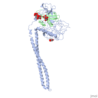
1jch
From Proteopedia
(Difference between revisions)
| Line 3: | Line 3: | ||
<StructureSection load='1jch' size='340' side='right'caption='[[1jch]], [[Resolution|resolution]] 3.02Å' scene=''> | <StructureSection load='1jch' size='340' side='right'caption='[[1jch]], [[Resolution|resolution]] 3.02Å' scene=''> | ||
== Structural highlights == | == Structural highlights == | ||
| - | <table><tr><td colspan='2'>[[1jch]] is a 4 chain structure with sequence from [https://en.wikipedia.org/wiki/ | + | <table><tr><td colspan='2'>[[1jch]] is a 4 chain structure with sequence from [https://en.wikipedia.org/wiki/Escherichia_coli_str._K-12_substr._W3110 Escherichia coli str. K-12 substr. W3110]. Full crystallographic information is available from [http://oca.weizmann.ac.il/oca-bin/ocashort?id=1JCH OCA]. For a <b>guided tour on the structure components</b> use [https://proteopedia.org/fgij/fg.htm?mol=1JCH FirstGlance]. <br> |
| - | </td></tr><tr id=' | + | </td></tr><tr id='method'><td class="sblockLbl"><b>[[Empirical_models|Method:]]</b></td><td class="sblockDat" id="methodDat">X-ray diffraction, [[Resolution|Resolution]] 3.02Å</td></tr> |
| - | <tr id=' | + | <tr id='ligand'><td class="sblockLbl"><b>[[Ligand|Ligands:]]</b></td><td class="sblockDat" id="ligandDat"><scene name='pdbligand=CIT:CITRIC+ACID'>CIT</scene>, <scene name='pdbligand=GOL:GLYCEROL'>GOL</scene></td></tr> |
<tr id='resources'><td class="sblockLbl"><b>Resources:</b></td><td class="sblockDat"><span class='plainlinks'>[https://proteopedia.org/fgij/fg.htm?mol=1jch FirstGlance], [http://oca.weizmann.ac.il/oca-bin/ocaids?id=1jch OCA], [https://pdbe.org/1jch PDBe], [https://www.rcsb.org/pdb/explore.do?structureId=1jch RCSB], [https://www.ebi.ac.uk/pdbsum/1jch PDBsum], [https://prosat.h-its.org/prosat/prosatexe?pdbcode=1jch ProSAT]</span></td></tr> | <tr id='resources'><td class="sblockLbl"><b>Resources:</b></td><td class="sblockDat"><span class='plainlinks'>[https://proteopedia.org/fgij/fg.htm?mol=1jch FirstGlance], [http://oca.weizmann.ac.il/oca-bin/ocaids?id=1jch OCA], [https://pdbe.org/1jch PDBe], [https://www.rcsb.org/pdb/explore.do?structureId=1jch RCSB], [https://www.ebi.ac.uk/pdbsum/1jch PDBsum], [https://prosat.h-its.org/prosat/prosatexe?pdbcode=1jch ProSAT]</span></td></tr> | ||
</table> | </table> | ||
== Function == | == Function == | ||
| - | + | [https://www.uniprot.org/uniprot/CEA3_ECOLX CEA3_ECOLX] Inactivates ribosomes by hydrolyzing 16S RNA in 30S ribosomes at a specific site. Colicins are polypeptide toxins produced by and active against E.coli and closely related bacteria. | |
== Evolutionary Conservation == | == Evolutionary Conservation == | ||
[[Image:Consurf_key_small.gif|200px|right]] | [[Image:Consurf_key_small.gif|200px|right]] | ||
| Line 20: | Line 20: | ||
</jmol>, as determined by [http://consurfdb.tau.ac.il/ ConSurfDB]. You may read the [[Conservation%2C_Evolutionary|explanation]] of the method and the full data available from [http://bental.tau.ac.il/new_ConSurfDB/main_output.php?pdb_ID=1jch ConSurf]. | </jmol>, as determined by [http://consurfdb.tau.ac.il/ ConSurfDB]. You may read the [[Conservation%2C_Evolutionary|explanation]] of the method and the full data available from [http://bental.tau.ac.il/new_ConSurfDB/main_output.php?pdb_ID=1jch ConSurf]. | ||
<div style="clear:both"></div> | <div style="clear:both"></div> | ||
| - | <div style="background-color:#fffaf0;"> | ||
| - | == Publication Abstract from PubMed == | ||
| - | Colicins kill E. coli by a process that involves binding to a surface receptor, entering the cell, and, finally, intoxicating it. The lethal action of colicin E3 is a specific cleavage in the ribosomal decoding A site. The crystal structure of colicin E3, reported here in a binary complex with its immunity protein (IP), reveals a Y-shaped molecule with the receptor binding domain forming a 100 A long stalk and the two globular heads of the translocation domain (T) and the catalytic domain (C) comprising the two arms. Active site residues are D510, H513, E517, and R545. IP is buried between T and C. Rather than blocking the active site, IP prevents access of the active site to the ribosome. | ||
| - | |||
| - | Crystal structure of colicin E3: implications for cell entry and ribosome inactivation.,Soelaiman S, Jakes K, Wu N, Li C, Shoham M Mol Cell. 2001 Nov;8(5):1053-62. PMID:11741540<ref>PMID:11741540</ref> | ||
| - | |||
| - | From MEDLINE®/PubMed®, a database of the U.S. National Library of Medicine.<br> | ||
| - | </div> | ||
| - | <div class="pdbe-citations 1jch" style="background-color:#fffaf0;"></div> | ||
==See Also== | ==See Also== | ||
*[[Colicin 3D structures|Colicin 3D structures]] | *[[Colicin 3D structures|Colicin 3D structures]] | ||
*[[Colicin immunity protein 3D structures|Colicin immunity protein 3D structures]] | *[[Colicin immunity protein 3D structures|Colicin immunity protein 3D structures]] | ||
| - | == References == | ||
| - | <references/> | ||
__TOC__ | __TOC__ | ||
</StructureSection> | </StructureSection> | ||
| - | [[Category: | + | [[Category: Escherichia coli str. K-12 substr. W3110]] |
[[Category: Large Structures]] | [[Category: Large Structures]] | ||
| - | [[Category: Jakes | + | [[Category: Jakes K]] |
| - | [[Category: Li | + | [[Category: Li C]] |
| - | [[Category: Shoham | + | [[Category: Shoham M]] |
| - | [[Category: Soelaiman | + | [[Category: Soelaiman S]] |
| - | [[Category: Wu | + | [[Category: Wu N]] |
| - | + | ||
| - | + | ||
| - | + | ||
| - | + | ||
| - | + | ||
Current revision
Crystal Structure of Colicin E3 in Complex with its Immunity Protein
| |||||||||||