We apologize for Proteopedia being slow to respond. For the past two years, a new implementation of Proteopedia has been being built. Soon, it will replace this 18-year old system. All existing content will be moved to the new system at a date that will be announced here.
1qav
From Proteopedia
(Difference between revisions)
| Line 3: | Line 3: | ||
<StructureSection load='1qav' size='340' side='right'caption='[[1qav]], [[Resolution|resolution]] 1.90Å' scene=''> | <StructureSection load='1qav' size='340' side='right'caption='[[1qav]], [[Resolution|resolution]] 1.90Å' scene=''> | ||
== Structural highlights == | == Structural highlights == | ||
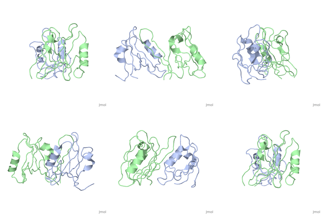
| - | <table><tr><td colspan='2'>[[1qav]] is a 2 chain structure with sequence from [https://en.wikipedia.org/wiki/ | + | <table><tr><td colspan='2'>[[1qav]] is a 2 chain structure with sequence from [https://en.wikipedia.org/wiki/Mus_musculus Mus musculus] and [https://en.wikipedia.org/wiki/Rattus_norvegicus Rattus norvegicus]. Full crystallographic information is available from [http://oca.weizmann.ac.il/oca-bin/ocashort?id=1QAV OCA]. For a <b>guided tour on the structure components</b> use [https://proteopedia.org/fgij/fg.htm?mol=1QAV FirstGlance]. <br> |
| - | </td></tr><tr id='resources'><td class="sblockLbl"><b>Resources:</b></td><td class="sblockDat"><span class='plainlinks'>[https://proteopedia.org/fgij/fg.htm?mol=1qav FirstGlance], [http://oca.weizmann.ac.il/oca-bin/ocaids?id=1qav OCA], [https://pdbe.org/1qav PDBe], [https://www.rcsb.org/pdb/explore.do?structureId=1qav RCSB], [https://www.ebi.ac.uk/pdbsum/1qav PDBsum], [https://prosat.h-its.org/prosat/prosatexe?pdbcode=1qav ProSAT]</span></td></tr> | + | </td></tr><tr id='method'><td class="sblockLbl"><b>[[Empirical_models|Method:]]</b></td><td class="sblockDat" id="methodDat">X-ray diffraction, [[Resolution|Resolution]] 1.9Å</td></tr> |
| + | <tr id='resources'><td class="sblockLbl"><b>Resources:</b></td><td class="sblockDat"><span class='plainlinks'>[https://proteopedia.org/fgij/fg.htm?mol=1qav FirstGlance], [http://oca.weizmann.ac.il/oca-bin/ocaids?id=1qav OCA], [https://pdbe.org/1qav PDBe], [https://www.rcsb.org/pdb/explore.do?structureId=1qav RCSB], [https://www.ebi.ac.uk/pdbsum/1qav PDBsum], [https://prosat.h-its.org/prosat/prosatexe?pdbcode=1qav ProSAT]</span></td></tr> | ||
</table> | </table> | ||
== Function == | == Function == | ||
| - | + | [https://www.uniprot.org/uniprot/SNTA1_MOUSE SNTA1_MOUSE] Adapter protein that binds to and probably organizes the subcellular localization of a variety of membrane proteins. May link various receptors to the actin cytoskeleton and the extracellular matrix via the dystrophin glycoprotein complex. Plays an important role in synapse formation and in the organization of UTRN and acetylcholine receptors at the neuromuscular synapse. Binds to phosphatidylinositol 4,5-bisphosphate. | |
== Evolutionary Conservation == | == Evolutionary Conservation == | ||
[[Image:Consurf_key_small.gif|200px|right]] | [[Image:Consurf_key_small.gif|200px|right]] | ||
| Line 18: | Line 19: | ||
</jmol>, as determined by [http://consurfdb.tau.ac.il/ ConSurfDB]. You may read the [[Conservation%2C_Evolutionary|explanation]] of the method and the full data available from [http://bental.tau.ac.il/new_ConSurfDB/main_output.php?pdb_ID=1qav ConSurf]. | </jmol>, as determined by [http://consurfdb.tau.ac.il/ ConSurfDB]. You may read the [[Conservation%2C_Evolutionary|explanation]] of the method and the full data available from [http://bental.tau.ac.il/new_ConSurfDB/main_output.php?pdb_ID=1qav ConSurf]. | ||
<div style="clear:both"></div> | <div style="clear:both"></div> | ||
| - | <div style="background-color:#fffaf0;"> | ||
| - | == Publication Abstract from PubMed == | ||
| - | The PDZ protein interaction domain of neuronal nitric oxide synthase (nNOS) can heterodimerize with the PDZ domains of postsynaptic density protein 95 and syntrophin through interactions that are not mediated by recognition of a typical carboxyl-terminal motif. The nNOS-syntrophin PDZ complex structure revealed that the domains interact in an unusual linear head-to-tail arrangement. The nNOS PDZ domain has two opposite interaction surfaces-one face has the canonical peptide binding groove, whereas the other has a beta-hairpin "finger." This nNOS beta finger docks in the syntrophin peptide binding groove, mimicking a peptide ligand, except that a sharp beta turn replaces the normally required carboxyl terminus. This structure explains how PDZ domains can participate in diverse interaction modes to assemble protein networks. | ||
| - | |||
| - | Unexpected modes of PDZ domain scaffolding revealed by structure of nNOS-syntrophin complex.,Hillier BJ, Christopherson KS, Prehoda KE, Bredt DS, Lim WA Science. 1999 Apr 30;284(5415):812-5. PMID:10221915<ref>PMID:10221915</ref> | ||
| - | |||
| - | From MEDLINE®/PubMed®, a database of the U.S. National Library of Medicine.<br> | ||
| - | </div> | ||
| - | <div class="pdbe-citations 1qav" style="background-color:#fffaf0;"></div> | ||
==See Also== | ==See Also== | ||
*[[Nitric Oxide Synthase 3D structures|Nitric Oxide Synthase 3D structures]] | *[[Nitric Oxide Synthase 3D structures|Nitric Oxide Synthase 3D structures]] | ||
*[[Syntrophin|Syntrophin]] | *[[Syntrophin|Syntrophin]] | ||
| - | == References == | ||
| - | <references/> | ||
__TOC__ | __TOC__ | ||
</StructureSection> | </StructureSection> | ||
| - | [[Category: Buffalo rat]] | ||
[[Category: Large Structures]] | [[Category: Large Structures]] | ||
| - | [[Category: | + | [[Category: Mus musculus]] |
| - | [[Category: | + | [[Category: Rattus norvegicus]] |
| - | [[Category: | + | [[Category: Bredt DS]] |
| - | [[Category: | + | [[Category: Christopherson KS]] |
| - | [[Category: | + | [[Category: Hillier BJ]] |
| - | [[Category: | + | [[Category: Lim WA]] |
| - | [[Category: | + | [[Category: Prehoda KE]] |
| - | + | ||
| - | + | ||
Current revision
Unexpected Modes of PDZ Domain Scaffolding Revealed by Structure of NNOS-Syntrophin Complex
| |||||||||||


