We apologize for Proteopedia being slow to respond. For the past two years, a new implementation of Proteopedia has been being built. Soon, it will replace this 18-year old system. All existing content will be moved to the new system at a date that will be announced here.
1sh6
From Proteopedia
(Difference between revisions)
| Line 3: | Line 3: | ||
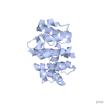
<StructureSection load='1sh6' size='340' side='right'caption='[[1sh6]], [[Resolution|resolution]] 2.00Å' scene=''> | <StructureSection load='1sh6' size='340' side='right'caption='[[1sh6]], [[Resolution|resolution]] 2.00Å' scene=''> | ||
== Structural highlights == | == Structural highlights == | ||
| - | <table><tr><td colspan='2'>[[1sh6]] is a 1 chain structure with sequence from [https://en.wikipedia.org/wiki/ | + | <table><tr><td colspan='2'>[[1sh6]] is a 1 chain structure with sequence from [https://en.wikipedia.org/wiki/Mus_musculus Mus musculus]. Full crystallographic information is available from [http://oca.weizmann.ac.il/oca-bin/ocashort?id=1SH6 OCA]. For a <b>guided tour on the structure components</b> use [https://proteopedia.org/fgij/fg.htm?mol=1SH6 FirstGlance]. <br> |
| - | </td></tr><tr id=' | + | </td></tr><tr id='method'><td class="sblockLbl"><b>[[Empirical_models|Method:]]</b></td><td class="sblockDat" id="methodDat">X-ray diffraction, [[Resolution|Resolution]] 2Å</td></tr> |
<tr id='resources'><td class="sblockLbl"><b>Resources:</b></td><td class="sblockDat"><span class='plainlinks'>[https://proteopedia.org/fgij/fg.htm?mol=1sh6 FirstGlance], [http://oca.weizmann.ac.il/oca-bin/ocaids?id=1sh6 OCA], [https://pdbe.org/1sh6 PDBe], [https://www.rcsb.org/pdb/explore.do?structureId=1sh6 RCSB], [https://www.ebi.ac.uk/pdbsum/1sh6 PDBsum], [https://prosat.h-its.org/prosat/prosatexe?pdbcode=1sh6 ProSAT]</span></td></tr> | <tr id='resources'><td class="sblockLbl"><b>Resources:</b></td><td class="sblockDat"><span class='plainlinks'>[https://proteopedia.org/fgij/fg.htm?mol=1sh6 FirstGlance], [http://oca.weizmann.ac.il/oca-bin/ocaids?id=1sh6 OCA], [https://pdbe.org/1sh6 PDBe], [https://www.rcsb.org/pdb/explore.do?structureId=1sh6 RCSB], [https://www.ebi.ac.uk/pdbsum/1sh6 PDBsum], [https://prosat.h-its.org/prosat/prosatexe?pdbcode=1sh6 ProSAT]</span></td></tr> | ||
</table> | </table> | ||
== Function == | == Function == | ||
| - | + | [https://www.uniprot.org/uniprot/PLEC_MOUSE PLEC_MOUSE] Interlinks intermediate filaments with microtubules and microfilaments and anchors intermediate filaments to desmosomes or hemidesmosomes. May be involved not only in the cross-linking and stabilization of cytoskeletal intermediate filaments network, but also in the regulation of their dynamics. | |
== Evolutionary Conservation == | == Evolutionary Conservation == | ||
[[Image:Consurf_key_small.gif|200px|right]] | [[Image:Consurf_key_small.gif|200px|right]] | ||
| Line 19: | Line 19: | ||
</jmol>, as determined by [http://consurfdb.tau.ac.il/ ConSurfDB]. You may read the [[Conservation%2C_Evolutionary|explanation]] of the method and the full data available from [http://bental.tau.ac.il/new_ConSurfDB/main_output.php?pdb_ID=1sh6 ConSurf]. | </jmol>, as determined by [http://consurfdb.tau.ac.il/ ConSurfDB]. You may read the [[Conservation%2C_Evolutionary|explanation]] of the method and the full data available from [http://bental.tau.ac.il/new_ConSurfDB/main_output.php?pdb_ID=1sh6 ConSurf]. | ||
<div style="clear:both"></div> | <div style="clear:both"></div> | ||
| - | <div style="background-color:#fffaf0;"> | ||
| - | == Publication Abstract from PubMed == | ||
| - | Plectin, a large and widely expressed cytolinker protein, is composed of several subdomains that harbor binding sites for a variety of different interaction partners. A canonical actin-binding domain (ABD) comprising two calponin homology domains (CH1 and CH2) is located in proximity to its amino terminus. However, the ABD of plectin is unique among actin-binding proteins as it is expressed in the form of distinct, plectin isoform-specific versions. We have determined the three-dimensional structure of two distinct crystalline forms of one of its ABD versions (pleABD/2alpha) from mouse, to a resolution of 1.95 and 2.0 A. Comparison of pleABD/2alpha with the ABDs of fimbrin and utrophin revealed structural similarity between plectin and fimbrin, although the proteins share only low sequence identity. In fact, pleABD/2alpha has been found to have the same compact fold as the human plectin ABD and the fimbrin ABD, differing from the open conformation described for the ABDs of utrophin and dystrophin. Plectin harbors a specific binding site for intermediate filaments of various types within its carboxy-terminal R5 repeat domain. Our experiments revealed an additional vimentin-binding site of plectin, residing within the CH1 subdomain of its ABD. We show that vimentin binds to this site via the amino-terminal part of its rod domain. This additional amino-terminal intermediate filament protein binding site of plectin may have a function in intermediate filament dynamics and assembly, rather than in linking and stabilizing intermediate filament networks. | ||
| - | |||
| - | Actin-binding domain of mouse plectin. Crystal structure and binding to vimentin.,Sevcik J, Urbanikova L, Kost'an J, Janda L, Wiche G Eur J Biochem. 2004 May;271(10):1873-84. PMID:15128297<ref>PMID:15128297</ref> | ||
| - | |||
| - | From MEDLINE®/PubMed®, a database of the U.S. National Library of Medicine.<br> | ||
| - | </div> | ||
| - | <div class="pdbe-citations 1sh6" style="background-color:#fffaf0;"></div> | ||
==See Also== | ==See Also== | ||
*[[Plectin|Plectin]] | *[[Plectin|Plectin]] | ||
| - | == References == | ||
| - | <references/> | ||
__TOC__ | __TOC__ | ||
</StructureSection> | </StructureSection> | ||
[[Category: Large Structures]] | [[Category: Large Structures]] | ||
| - | [[Category: | + | [[Category: Mus musculus]] |
| - | [[Category: Sevcik | + | [[Category: Sevcik J]] |
| - | [[Category: Urbanikova | + | [[Category: Urbanikova L]] |
| - | + | ||
| - | + | ||
| - | + | ||
| - | + | ||
Current revision
Crystal structure of actin-binding domain of mouse plectin
| |||||||||||