This old version of Proteopedia is provided for student assignments while the new version is undergoing repairs. Content and edits done in this old version of Proteopedia after March 1, 2026 will eventually be lost when it is retired in about June of 2026.
Apply for new accounts at the new Proteopedia. Your logins will work in both the old and new versions.
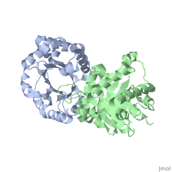
1ypi
From Proteopedia
(Difference between revisions)
| Line 3: | Line 3: | ||
<StructureSection load='1ypi' size='340' side='right'caption='[[1ypi]], [[Resolution|resolution]] 1.90Å' scene=''> | <StructureSection load='1ypi' size='340' side='right'caption='[[1ypi]], [[Resolution|resolution]] 1.90Å' scene=''> | ||
== Structural highlights == | == Structural highlights == | ||
| - | <table><tr><td colspan='2'>[[1ypi]] is a 2 chain structure with sequence from [https://en.wikipedia.org/wiki/ | + | <table><tr><td colspan='2'>[[1ypi]] is a 2 chain structure with sequence from [https://en.wikipedia.org/wiki/Saccharomyces_cerevisiae Saccharomyces cerevisiae]. Full crystallographic information is available from [http://oca.weizmann.ac.il/oca-bin/ocashort?id=1YPI OCA]. For a <b>guided tour on the structure components</b> use [https://proteopedia.org/fgij/fg.htm?mol=1YPI FirstGlance]. <br> |
| - | </td></tr><tr id=' | + | </td></tr><tr id='method'><td class="sblockLbl"><b>[[Empirical_models|Method:]]</b></td><td class="sblockDat" id="methodDat">X-ray diffraction, [[Resolution|Resolution]] 1.9Å</td></tr> |
<tr id='resources'><td class="sblockLbl"><b>Resources:</b></td><td class="sblockDat"><span class='plainlinks'>[https://proteopedia.org/fgij/fg.htm?mol=1ypi FirstGlance], [http://oca.weizmann.ac.il/oca-bin/ocaids?id=1ypi OCA], [https://pdbe.org/1ypi PDBe], [https://www.rcsb.org/pdb/explore.do?structureId=1ypi RCSB], [https://www.ebi.ac.uk/pdbsum/1ypi PDBsum], [https://prosat.h-its.org/prosat/prosatexe?pdbcode=1ypi ProSAT]</span></td></tr> | <tr id='resources'><td class="sblockLbl"><b>Resources:</b></td><td class="sblockDat"><span class='plainlinks'>[https://proteopedia.org/fgij/fg.htm?mol=1ypi FirstGlance], [http://oca.weizmann.ac.il/oca-bin/ocaids?id=1ypi OCA], [https://pdbe.org/1ypi PDBe], [https://www.rcsb.org/pdb/explore.do?structureId=1ypi RCSB], [https://www.ebi.ac.uk/pdbsum/1ypi PDBsum], [https://prosat.h-its.org/prosat/prosatexe?pdbcode=1ypi ProSAT]</span></td></tr> | ||
</table> | </table> | ||
| + | == Function == | ||
| + | [https://www.uniprot.org/uniprot/TPIS_YEAST TPIS_YEAST] | ||
== Evolutionary Conservation == | == Evolutionary Conservation == | ||
[[Image:Consurf_key_small.gif|200px|right]] | [[Image:Consurf_key_small.gif|200px|right]] | ||
| Line 17: | Line 19: | ||
</jmol>, as determined by [http://consurfdb.tau.ac.il/ ConSurfDB]. You may read the [[Conservation%2C_Evolutionary|explanation]] of the method and the full data available from [http://bental.tau.ac.il/new_ConSurfDB/main_output.php?pdb_ID=1ypi ConSurf]. | </jmol>, as determined by [http://consurfdb.tau.ac.il/ ConSurfDB]. You may read the [[Conservation%2C_Evolutionary|explanation]] of the method and the full data available from [http://bental.tau.ac.il/new_ConSurfDB/main_output.php?pdb_ID=1ypi ConSurf]. | ||
<div style="clear:both"></div> | <div style="clear:both"></div> | ||
| - | <div style="background-color:#fffaf0;"> | ||
| - | == Publication Abstract from PubMed == | ||
| - | The structure of yeast triosephosphate isomerase (TIM) has been solved at 3.0-A resolution and refined at 1.9-A resolution to an R factor of 21.0%. The final model consists of all non-hydrogen atoms in the polypeptide chain and 119 water molecules, a number of which are found in the interior of the protein. The structure of the active site clearly indicates that the carboxylate of the catalytic base, Glu 165, is involved in a hydrogen-bonding interaction with the hydroxyl of Ser 96. In addition, the interactions of the other active site residues, Lys 12 and His 95, are also discussed. For the first time in any TIM structure, the "flexible loop" has well-defined density; the conformation of the loop in this structure is stabilized by a crystal contact. Analysis of the subunit interface of this dimeric enzyme hints at the source of the specificity of one subunit for another and allows us to estimate an association constant of 10(14)-10(16) M-1 for the two monomers. The analysis also suggests that the interface may be a particularly good target for drug design. The conserved positions (20%) among sequences from 13 sources ranging on the evolutionary scale from Escherichia coli to humans reveal the intense pressure to maintain the active site structure. | ||
| - | |||
| - | Structure of yeast triosephosphate isomerase at 1.9-A resolution.,Lolis E, Alber T, Davenport RC, Rose D, Hartman FC, Petsko GA Biochemistry. 1990 Jul 17;29(28):6609-18. PMID:2204417<ref>PMID:2204417</ref> | ||
| - | |||
| - | From MEDLINE®/PubMed®, a database of the U.S. National Library of Medicine.<br> | ||
| - | </div> | ||
| - | <div class="pdbe-citations 1ypi" style="background-color:#fffaf0;"></div> | ||
==See Also== | ==See Also== | ||
*[[Triose phosphate isomerase 3D structures|Triose phosphate isomerase 3D structures]] | *[[Triose phosphate isomerase 3D structures|Triose phosphate isomerase 3D structures]] | ||
| - | == References == | ||
| - | <references/> | ||
__TOC__ | __TOC__ | ||
</StructureSection> | </StructureSection> | ||
| - | [[Category: Atcc 18824]] | ||
[[Category: Large Structures]] | [[Category: Large Structures]] | ||
| - | [[Category: | + | [[Category: Saccharomyces cerevisiae]] |
| - | [[Category: Alber | + | [[Category: Alber T]] |
| - | [[Category: Lolis | + | [[Category: Lolis E]] |
| - | [[Category: Petsko | + | [[Category: Petsko GA]] |
Current revision
STRUCTURE OF YEAST TRIOSEPHOSPHATE ISOMERASE AT 1.9-ANGSTROMS RESOLUTION
| |||||||||||