We apologize for Proteopedia being slow to respond. For the past two years, a new implementation of Proteopedia has been being built. Soon, it will replace this 18-year old system. All existing content will be moved to the new system at a date that will be announced here.
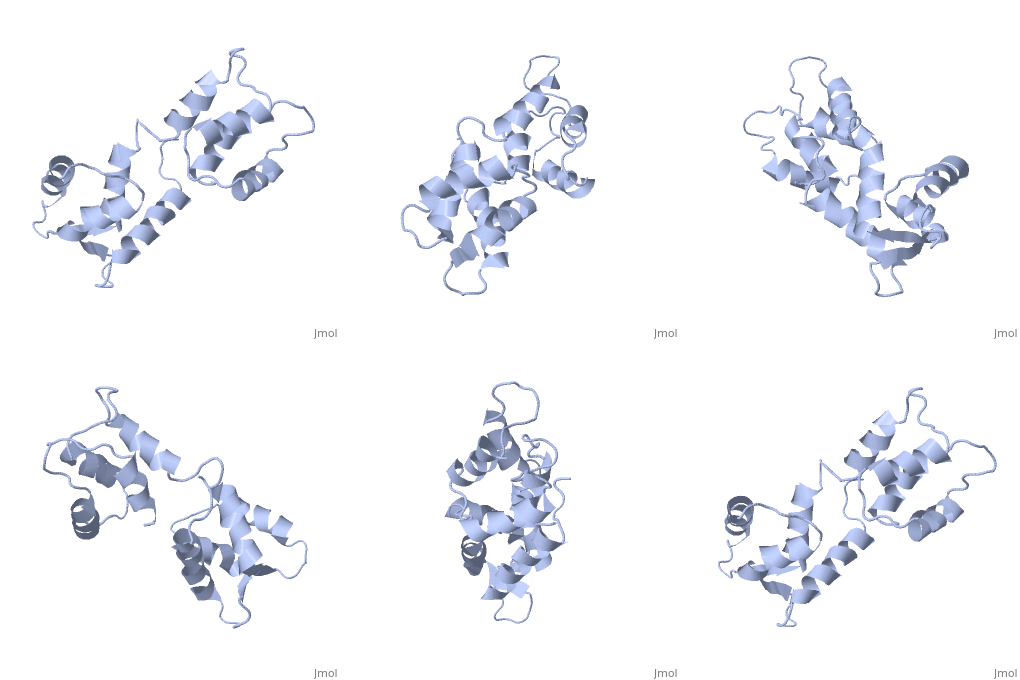
1cfc
From Proteopedia
(Difference between revisions)
| Line 1: | Line 1: | ||
==CALCIUM-FREE CALMODULIN== | ==CALCIUM-FREE CALMODULIN== | ||
| - | <StructureSection load='1cfc' size='340' side='right'caption='[[1cfc | + | <StructureSection load='1cfc' size='340' side='right'caption='[[1cfc]]' scene=''> |
== Structural highlights == | == Structural highlights == | ||
<table><tr><td colspan='2'>[[1cfc]] is a 1 chain structure with sequence from [https://en.wikipedia.org/wiki/Xenopus_laevis Xenopus laevis]. Full experimental information is available from [http://oca.weizmann.ac.il/oca-bin/ocashort?id=1CFC OCA]. For a <b>guided tour on the structure components</b> use [https://proteopedia.org/fgij/fg.htm?mol=1CFC FirstGlance]. <br> | <table><tr><td colspan='2'>[[1cfc]] is a 1 chain structure with sequence from [https://en.wikipedia.org/wiki/Xenopus_laevis Xenopus laevis]. Full experimental information is available from [http://oca.weizmann.ac.il/oca-bin/ocashort?id=1CFC OCA]. For a <b>guided tour on the structure components</b> use [https://proteopedia.org/fgij/fg.htm?mol=1CFC FirstGlance]. <br> | ||
| - | </td></tr><tr id=' | + | </td></tr><tr id='method'><td class="sblockLbl"><b>[[Empirical_models|Method:]]</b></td><td class="sblockDat" id="methodDat">Solution NMR</td></tr> |
<tr id='resources'><td class="sblockLbl"><b>Resources:</b></td><td class="sblockDat"><span class='plainlinks'>[https://proteopedia.org/fgij/fg.htm?mol=1cfc FirstGlance], [http://oca.weizmann.ac.il/oca-bin/ocaids?id=1cfc OCA], [https://pdbe.org/1cfc PDBe], [https://www.rcsb.org/pdb/explore.do?structureId=1cfc RCSB], [https://www.ebi.ac.uk/pdbsum/1cfc PDBsum], [https://prosat.h-its.org/prosat/prosatexe?pdbcode=1cfc ProSAT]</span></td></tr> | <tr id='resources'><td class="sblockLbl"><b>Resources:</b></td><td class="sblockDat"><span class='plainlinks'>[https://proteopedia.org/fgij/fg.htm?mol=1cfc FirstGlance], [http://oca.weizmann.ac.il/oca-bin/ocaids?id=1cfc OCA], [https://pdbe.org/1cfc PDBe], [https://www.rcsb.org/pdb/explore.do?structureId=1cfc RCSB], [https://www.ebi.ac.uk/pdbsum/1cfc PDBsum], [https://prosat.h-its.org/prosat/prosatexe?pdbcode=1cfc ProSAT]</span></td></tr> | ||
</table> | </table> | ||
== Function == | == Function == | ||
| - | + | [https://www.uniprot.org/uniprot/CALM1_XENLA CALM1_XENLA] Calmodulin mediates the control of a large number of enzymes, ion channels and other proteins by Ca(2+). Among the enzymes to be stimulated by the calmodulin-Ca(2+) complex are a number of protein kinases and phosphatases. | |
== Evolutionary Conservation == | == Evolutionary Conservation == | ||
[[Image:Consurf_key_small.gif|200px|right]] | [[Image:Consurf_key_small.gif|200px|right]] | ||
| Line 19: | Line 19: | ||
</jmol>, as determined by [http://consurfdb.tau.ac.il/ ConSurfDB]. You may read the [[Conservation%2C_Evolutionary|explanation]] of the method and the full data available from [http://bental.tau.ac.il/new_ConSurfDB/main_output.php?pdb_ID=1cfc ConSurf]. | </jmol>, as determined by [http://consurfdb.tau.ac.il/ ConSurfDB]. You may read the [[Conservation%2C_Evolutionary|explanation]] of the method and the full data available from [http://bental.tau.ac.il/new_ConSurfDB/main_output.php?pdb_ID=1cfc ConSurf]. | ||
<div style="clear:both"></div> | <div style="clear:both"></div> | ||
| - | <div style="background-color:#fffaf0;"> | ||
| - | == Publication Abstract from PubMed == | ||
| - | The three-dimensional structure of calmodulin in the absence of Ca2+ has been determined by three- and four-dimensional heteronuclear NMR experiments, including ROE, isotope-filtering combined with reverse labelling, and measurement of more than 700 three-bond J-couplings. In analogy with the Ca(2+)-ligated state of this protein, it consists of two small globular domains separated by a flexible linker, with no stable, direct contacts between the two domains. In the absence of Ca2+, the four helices in each of the two globular domains form a highly twisted bundle, capped by a short anti-parallel beta-sheet. This arrangement is qualitatively similar to that observed in the crystal structure of the Ca(2+)-free N-terminal domain of troponin C. | ||
| - | |||
| - | Solution structure of calcium-free calmodulin.,Kuboniwa H, Tjandra N, Grzesiek S, Ren H, Klee CB, Bax A Nat Struct Biol. 1995 Sep;2(9):768-76. PMID:7552748<ref>PMID:7552748</ref> | ||
| - | |||
| - | From MEDLINE®/PubMed®, a database of the U.S. National Library of Medicine.<br> | ||
| - | </div> | ||
| - | <div class="pdbe-citations 1cfc" style="background-color:#fffaf0;"></div> | ||
==See Also== | ==See Also== | ||
*[[Calmodulin 3D structures|Calmodulin 3D structures]] | *[[Calmodulin 3D structures|Calmodulin 3D structures]] | ||
*[[Hydrogen in macromolecular models|Hydrogen in macromolecular models]] | *[[Hydrogen in macromolecular models|Hydrogen in macromolecular models]] | ||
| - | == References == | ||
| - | <references/> | ||
__TOC__ | __TOC__ | ||
</StructureSection> | </StructureSection> | ||
[[Category: Large Structures]] | [[Category: Large Structures]] | ||
[[Category: Xenopus laevis]] | [[Category: Xenopus laevis]] | ||
| - | [[Category: Bax | + | [[Category: Bax A]] |
| - | [[Category: Grzesiek | + | [[Category: Grzesiek S]] |
| - | [[Category: Klee | + | [[Category: Klee CB]] |
| - | [[Category: Kuboniwa | + | [[Category: Kuboniwa H]] |
| - | [[Category: Ren | + | [[Category: Ren H]] |
| - | [[Category: Tjandra | + | [[Category: Tjandra N]] |
| - | + | ||
Revision as of 15:39, 13 March 2024
CALCIUM-FREE CALMODULIN
| |||||||||||
Categories: Large Structures | Xenopus laevis | Bax A | Grzesiek S | Klee CB | Kuboniwa H | Ren H | Tjandra N


