User:Chloe Tucker/Sandbox 1
From Proteopedia
(Difference between revisions)
| Line 14: | Line 14: | ||
== Structural highlights == | == Structural highlights == | ||
| - | <scene name='10/1038815/N-term_of_gip/23'>Binding site of GIP receptor</scene> | ||
This is a sample scene created with SAT to of the protein. You can make your own scenes on SAT starting from scratch or loading and editing one of these sample scenes. | This is a sample scene created with SAT to of the protein. You can make your own scenes on SAT starting from scratch or loading and editing one of these sample scenes. | ||
=== Binding/Active Site of GIPR with GIP === | === Binding/Active Site of GIPR with GIP === | ||
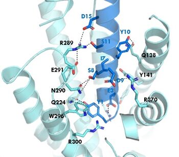
[[Image:GIP_hydrogen_bonds.jpg|350 px|right|thumb|Figure 1. Residue Interactions with GIP]] | [[Image:GIP_hydrogen_bonds.jpg|350 px|right|thumb|Figure 1. Residue Interactions with GIP]] | ||
| - | The <scene name='10/1038815/Overview/3'> | + | The <scene name='10/1038815/Overview/3'>binding site</scene> of GIP with the GIP receptor (GIPR) is where the N-term of GIP binds with the transmembrane domain of the GIP receptor. This first interaction formed with GIPR and the N-term of GIP is a hydrogen bond between TYR1 and GLU224. |
| - | The <scene name='10/1038815/Active_site/3'>residues</scene> within the active site are forming hydrogen bonds with each other and activating the G-protein to start | + | The <scene name='10/1038815/Active_site/3'>residues</scene> within the active site are forming hydrogen bonds with each other and activating the G-protein to start signaling. |
=== Binding/Active Site of GIPR with Tirzepatide === | === Binding/Active Site of GIPR with Tirzepatide === | ||
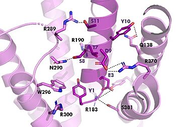
[[Image:TZ_hydrogen_bonds.jpg|350 px|right|thumb|Figure 2. Residue Interactions with Tirzepatide]] | [[Image:TZ_hydrogen_bonds.jpg|350 px|right|thumb|Figure 2. Residue Interactions with Tirzepatide]] | ||
Revision as of 13:18, 25 April 2024
GIP and GIP-R
| |||||||||||
References
- ↑ Hanson, R. M., Prilusky, J., Renjian, Z., Nakane, T. and Sussman, J. L. (2013), JSmol and the Next-Generation Web-Based Representation of 3D Molecular Structure as Applied to Proteopedia. Isr. J. Chem., 53:207-216. doi:http://dx.doi.org/10.1002/ijch.201300024
- ↑ Herraez A. Biomolecules in the computer: Jmol to the rescue. Biochem Mol Biol Educ. 2006 Jul;34(4):255-61. doi: 10.1002/bmb.2006.494034042644. PMID:21638687 doi:10.1002/bmb.2006.494034042644
- ↑ Sun B, Willard FS, Feng D, Alsina-Fernandez J, Chen Q, Vieth M, Ho JD, Showalter AD, Stutsman C, Ding L, Suter TM, Dunbar JD, Carpenter JW, Mohammed FA, Aihara E, Brown RA, Bueno AB, Emmerson PJ, Moyers JS, Kobilka TS, Coghlan MP, Kobilka BK, Sloop KW. Structural determinants of dual incretin receptor agonism by tirzepatide. Proc Natl Acad Sci U S A. 2022 Mar 29;119(13):e2116506119. PMID:35333651 doi:10.1073/pnas.2116506119
Student Contributors
- Chloe Tucker
- Mandy Bechman


