1dsn
From Proteopedia
(Difference between revisions)
| Line 15: | Line 15: | ||
<jmolCheckbox> | <jmolCheckbox> | ||
<scriptWhenChecked>; select protein; define ~consurf_to_do selected; consurf_initial_scene = true; script "/wiki/ConSurf/ds/1dsn_consurf.spt"</scriptWhenChecked> | <scriptWhenChecked>; select protein; define ~consurf_to_do selected; consurf_initial_scene = true; script "/wiki/ConSurf/ds/1dsn_consurf.spt"</scriptWhenChecked> | ||
| - | <scriptWhenUnchecked>script /wiki/extensions/Proteopedia/spt/ | + | <scriptWhenUnchecked>script /wiki/extensions/Proteopedia/spt/initialview03.spt</scriptWhenUnchecked> |
<text>to colour the structure by Evolutionary Conservation</text> | <text>to colour the structure by Evolutionary Conservation</text> | ||
</jmolCheckbox> | </jmolCheckbox> | ||
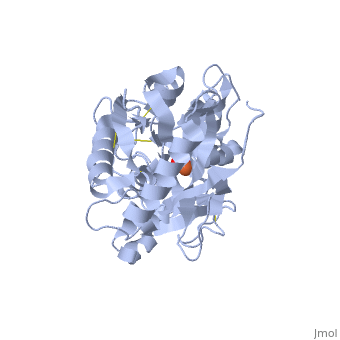
</jmol>, as determined by [http://consurfdb.tau.ac.il/ ConSurfDB]. You may read the [[Conservation%2C_Evolutionary|explanation]] of the method and the full data available from [http://bental.tau.ac.il/new_ConSurfDB/main_output.php?pdb_ID=1dsn ConSurf]. | </jmol>, as determined by [http://consurfdb.tau.ac.il/ ConSurfDB]. You may read the [[Conservation%2C_Evolutionary|explanation]] of the method and the full data available from [http://bental.tau.ac.il/new_ConSurfDB/main_output.php?pdb_ID=1dsn ConSurf]. | ||
<div style="clear:both"></div> | <div style="clear:both"></div> | ||
| + | <div style="background-color:#fffaf0;"> | ||
| + | == Publication Abstract from PubMed == | ||
| + | The crystal structure of a site-specific mutant of the N-terminal half-molecule of human lactoferrin, Lf(N), in which the iron ligand Asp60 has been mutated to Ser, has been determined at 2.05 A resolution in order to determine the effects of the mutation on iron binding and domain closure. Yellow monoclinic crystals of the D60S mutant, in its iron-bound form, were prepared, and have unit cell dimensions a = 110.2 A, b = 57.0 A, c = 55.2 A, beta = 97.6 degrees, space group C2, with one molecule of 333 residues in the asymmetric unit. The structure was determined by molecular replacement, using the wild-type Lf(N) as search model, and was refined by restrained least-squares methods. The final model, comprising 2451 protein atoms (from residues 2 to 315) one Fe3+ and one CO2-(3), and 107 water molecules, gives an R-factor of 0.175 for all data in the resolution range 20.0 to 2.05 A. The model conforms well with standard geometry, having root-mean-square deviations of 0.014 A and 1.2 degrees from standard bond lengths and angles. The structure of the D60S mutant deviates in two important respects from the parent Lf(N) molecule. At the mutation site the Ser side-chain neither binds to the iron atom nor makes any interdomain contact as the substituted Asp does; instead a water molecule fills the iron coordination site and participates in interdomain hydrogen bonding. The domain closure is also changed, with the D60S mutant having a more closed conformation. Consideration of crystal packing suggests that the altered domain closure is a genuine molecular property but both the iron coordination and interdomain contacts are consistent with weakened iron binding in the mutant. The implications for iron binding in transferrins generally are discussed. | ||
| + | |||
| + | Altered domain closure and iron binding in transferrins: the crystal structure of the Asp60Ser mutant of the amino-terminal half-molecule of human lactoferrin.,Faber HR, Bland T, Day CL, Norris GE, Tweedie JW, Baker EN J Mol Biol. 1996 Feb 23;256(2):352-63. PMID:8594202<ref>PMID:8594202</ref> | ||
| + | |||
| + | From MEDLINE®/PubMed®, a database of the U.S. National Library of Medicine.<br> | ||
| + | </div> | ||
| + | <div class="pdbe-citations 1dsn" style="background-color:#fffaf0;"></div> | ||
==See Also== | ==See Also== | ||
Current revision
D60S N-TERMINAL LOBE HUMAN LACTOFERRIN
| |||||||||||