We apologize for Proteopedia being slow to respond. For the past two years, a new implementation of Proteopedia has been being built. Soon, it will replace this 18-year old system. All existing content will be moved to the new system at a date that will be announced here.
1dtg
From Proteopedia
(Difference between revisions)
| Line 17: | Line 17: | ||
<jmolCheckbox> | <jmolCheckbox> | ||
<scriptWhenChecked>; select protein; define ~consurf_to_do selected; consurf_initial_scene = true; script "/wiki/ConSurf/dt/1dtg_consurf.spt"</scriptWhenChecked> | <scriptWhenChecked>; select protein; define ~consurf_to_do selected; consurf_initial_scene = true; script "/wiki/ConSurf/dt/1dtg_consurf.spt"</scriptWhenChecked> | ||
| - | <scriptWhenUnchecked>script /wiki/extensions/Proteopedia/spt/ | + | <scriptWhenUnchecked>script /wiki/extensions/Proteopedia/spt/initialview03.spt</scriptWhenUnchecked> |
<text>to colour the structure by Evolutionary Conservation</text> | <text>to colour the structure by Evolutionary Conservation</text> | ||
</jmolCheckbox> | </jmolCheckbox> | ||
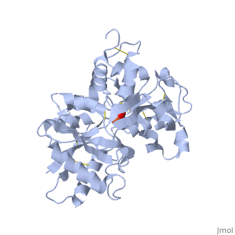
</jmol>, as determined by [http://consurfdb.tau.ac.il/ ConSurfDB]. You may read the [[Conservation%2C_Evolutionary|explanation]] of the method and the full data available from [http://bental.tau.ac.il/new_ConSurfDB/main_output.php?pdb_ID=1dtg ConSurf]. | </jmol>, as determined by [http://consurfdb.tau.ac.il/ ConSurfDB]. You may read the [[Conservation%2C_Evolutionary|explanation]] of the method and the full data available from [http://bental.tau.ac.il/new_ConSurfDB/main_output.php?pdb_ID=1dtg ConSurf]. | ||
<div style="clear:both"></div> | <div style="clear:both"></div> | ||
| + | <div style="background-color:#fffaf0;"> | ||
| + | == Publication Abstract from PubMed == | ||
| + | Serum transferrin is the major iron transport protein in humans. Its function depends on its ability to bind iron with very high affinity, yet to release this bound iron at the lower intracellular pH. Possible explanations for the release of iron from transferrin at low pH include protonation of a histidine ligand and the existence of a pH-sensitive "trigger" involving a hydrogen-bonded pair of lysines in the N-lobe of transferrin. We have determined the crystal structure of the His249Glu mutant of the N-lobe half-molecule of human transferrin and compared its iron-binding properties with those of the wild-type protein and other mutants. The crystal structure, determined at 2.4 A resolution (R-factor 19.8%, R(free) 29.4%), shows that Glu 249 is directly bound to iron, in place of the His ligand, and that a local movement of Lys 296 has broken the dilysine interaction. Despite the loss of this potentially pH-sensitive interaction, the H249E mutant is only slightly more acid-stable than wild-type and releases iron slightly faster. We conclude that the loss of the dilysine interaction does make the protein more acid stable but that this is counterbalanced by the replacement of a neutral ligand (His) by a negatively charged one (Glu), thus disrupting the electroneutrality of the binding site. | ||
| + | |||
| + | Mutation of the iron ligand His 249 to Glu in the N-lobe of human transferrin abolishes the dilysine "trigger" but does not significantly affect iron release.,MacGillivray RT, Bewley MC, Smith CA, He QY, Mason AB, Woodworth RC, Baker EN Biochemistry. 2000 Feb 15;39(6):1211-6. PMID:10684598<ref>PMID:10684598</ref> | ||
| + | |||
| + | From MEDLINE®/PubMed®, a database of the U.S. National Library of Medicine.<br> | ||
| + | </div> | ||
| + | <div class="pdbe-citations 1dtg" style="background-color:#fffaf0;"></div> | ||
==See Also== | ==See Also== | ||
Current revision
HUMAN TRANSFERRIN N-LOBE MUTANT H249E
| |||||||||||