We apologize for Proteopedia being slow to respond. For the past two years, a new implementation of Proteopedia has been being built. Soon, it will replace this 18-year old system. All existing content will be moved to the new system at a date that will be announced here.
1ls8
From Proteopedia
(Difference between revisions)
| Line 4: | Line 4: | ||
== Structural highlights == | == Structural highlights == | ||
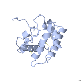
<table><tr><td colspan='2'>[[1ls8]] is a 1 chain structure with sequence from [https://en.wikipedia.org/wiki/Bombyx_mori Bombyx mori]. Full experimental information is available from [http://oca.weizmann.ac.il/oca-bin/ocashort?id=1LS8 OCA]. For a <b>guided tour on the structure components</b> use [https://proteopedia.org/fgij/fg.htm?mol=1LS8 FirstGlance]. <br> | <table><tr><td colspan='2'>[[1ls8]] is a 1 chain structure with sequence from [https://en.wikipedia.org/wiki/Bombyx_mori Bombyx mori]. Full experimental information is available from [http://oca.weizmann.ac.il/oca-bin/ocashort?id=1LS8 OCA]. For a <b>guided tour on the structure components</b> use [https://proteopedia.org/fgij/fg.htm?mol=1LS8 FirstGlance]. <br> | ||
| - | </td></tr><tr id='method'><td class="sblockLbl"><b>[[Empirical_models|Method:]]</b></td><td class="sblockDat" id="methodDat">Solution NMR</td></tr> | + | </td></tr><tr id='method'><td class="sblockLbl"><b>[[Empirical_models|Method:]]</b></td><td class="sblockDat" id="methodDat">Solution NMR, 20 models</td></tr> |
<tr id='resources'><td class="sblockLbl"><b>Resources:</b></td><td class="sblockDat"><span class='plainlinks'>[https://proteopedia.org/fgij/fg.htm?mol=1ls8 FirstGlance], [http://oca.weizmann.ac.il/oca-bin/ocaids?id=1ls8 OCA], [https://pdbe.org/1ls8 PDBe], [https://www.rcsb.org/pdb/explore.do?structureId=1ls8 RCSB], [https://www.ebi.ac.uk/pdbsum/1ls8 PDBsum], [https://prosat.h-its.org/prosat/prosatexe?pdbcode=1ls8 ProSAT]</span></td></tr> | <tr id='resources'><td class="sblockLbl"><b>Resources:</b></td><td class="sblockDat"><span class='plainlinks'>[https://proteopedia.org/fgij/fg.htm?mol=1ls8 FirstGlance], [http://oca.weizmann.ac.il/oca-bin/ocaids?id=1ls8 OCA], [https://pdbe.org/1ls8 PDBe], [https://www.rcsb.org/pdb/explore.do?structureId=1ls8 RCSB], [https://www.ebi.ac.uk/pdbsum/1ls8 PDBsum], [https://prosat.h-its.org/prosat/prosatexe?pdbcode=1ls8 ProSAT]</span></td></tr> | ||
</table> | </table> | ||
| Line 14: | Line 14: | ||
<jmolCheckbox> | <jmolCheckbox> | ||
<scriptWhenChecked>; select protein; define ~consurf_to_do selected; consurf_initial_scene = true; script "/wiki/ConSurf/ls/1ls8_consurf.spt"</scriptWhenChecked> | <scriptWhenChecked>; select protein; define ~consurf_to_do selected; consurf_initial_scene = true; script "/wiki/ConSurf/ls/1ls8_consurf.spt"</scriptWhenChecked> | ||
| - | <scriptWhenUnchecked>script /wiki/extensions/Proteopedia/spt/ | + | <scriptWhenUnchecked>script /wiki/extensions/Proteopedia/spt/initialview03.spt</scriptWhenUnchecked> |
<text>to colour the structure by Evolutionary Conservation</text> | <text>to colour the structure by Evolutionary Conservation</text> | ||
</jmolCheckbox> | </jmolCheckbox> | ||
</jmol>, as determined by [http://consurfdb.tau.ac.il/ ConSurfDB]. You may read the [[Conservation%2C_Evolutionary|explanation]] of the method and the full data available from [http://bental.tau.ac.il/new_ConSurfDB/main_output.php?pdb_ID=1ls8 ConSurf]. | </jmol>, as determined by [http://consurfdb.tau.ac.il/ ConSurfDB]. You may read the [[Conservation%2C_Evolutionary|explanation]] of the method and the full data available from [http://bental.tau.ac.il/new_ConSurfDB/main_output.php?pdb_ID=1ls8 ConSurf]. | ||
<div style="clear:both"></div> | <div style="clear:both"></div> | ||
| + | <div style="background-color:#fffaf0;"> | ||
| + | == Publication Abstract from PubMed == | ||
| + | The nuclear magnetic resonance structure of the unliganded pheromone-binding protein (PBP) from Bombyx mori at pH above 6.5, BmPBP(B), consists of seven helices with residues 3-8, 16-22, 29-32, 46-59, 70-79, 84-100, and 107-124, and contains the three disulfide bridges 19-54, 50-108, and 97-117. This polypeptide fold encloses a large hydrophobic cavity, with a sufficient volume to accommodate the natural ligand bombykol. The polypeptide folds in free BmPBP(B) and in crystals of a BmPBP-bombykol complex are nearly identical, indicating that the B-form of BmPBP in solution represents the active conformation for ligand binding. | ||
| + | |||
| + | NMR structure of the unliganded Bombyx mori pheromone-binding protein at physiological pH.,Lee D, Damberger FF, Peng G, Horst R, Guntert P, Nikonova L, Leal WS, Wuthrich K FEBS Lett. 2002 Nov 6;531(2):314-8. PMID:12417333<ref>PMID:12417333</ref> | ||
| + | |||
| + | From MEDLINE®/PubMed®, a database of the U.S. National Library of Medicine.<br> | ||
| + | </div> | ||
| + | <div class="pdbe-citations 1ls8" style="background-color:#fffaf0;"></div> | ||
==See Also== | ==See Also== | ||
*[[Odorant binding protein|Odorant binding protein]] | *[[Odorant binding protein|Odorant binding protein]] | ||
| + | == References == | ||
| + | <references/> | ||
__TOC__ | __TOC__ | ||
</StructureSection> | </StructureSection> | ||
Current revision
NMR structure of the unliganded Bombyx mori pheromone-binding protein at physiological pH
| |||||||||||

Categories: Bombyx mori | Large Structures | Damberger F | Guntert P | Horst R | Leal WS | Lee D | Wuthrich K
