User:Emily Hwang/Sandbox1
From Proteopedia
(Difference between revisions)
| Line 12: | Line 12: | ||
==Overall Topology== | ==Overall Topology== | ||
Leaf-branch compost bacterial cutinase, LCC, is a part of the [https://en.wikipedia.org/wiki/Serine_hydrolase serine hydrolase] family. It is a monomer that contains a total of 258 amino acid residues, with an amphipathic structure. It has a <scene name='10/1075191/Overall_topology/1'>secondary structure</scene> made up of alpha helices and beta turns, which correlate to the alpha-beta hydrolase family. The active site consists of a <scene name='10/1075191/Active_site_overall_topology/5'>catalytic triad</scene>, which is a common feature among serine hydrolases. Compared to other serine hydrolases and cutinases studied for plastic degradation, the LCC proved to be 33x more efficient. | Leaf-branch compost bacterial cutinase, LCC, is a part of the [https://en.wikipedia.org/wiki/Serine_hydrolase serine hydrolase] family. It is a monomer that contains a total of 258 amino acid residues, with an amphipathic structure. It has a <scene name='10/1075191/Overall_topology/1'>secondary structure</scene> made up of alpha helices and beta turns, which correlate to the alpha-beta hydrolase family. The active site consists of a <scene name='10/1075191/Active_site_overall_topology/5'>catalytic triad</scene>, which is a common feature among serine hydrolases. Compared to other serine hydrolases and cutinases studied for plastic degradation, the LCC proved to be 33x more efficient. | ||
| - | =Active Site= | + | ==Active Site== |
==Structure== | ==Structure== | ||
The active site contains a <scene name='10/1075193/Hydrophobic_binding_pocket/6'>hydrophobic binding pocket</scene> which makes [https://en.wikipedia.org/wiki/Pi-stacking aromatic pi-stacking] and [https://en.wikipedia.org/wiki/Van_der_Waals_force Van der Waals interactions] with the aromatic rings in the PET ligand. Add a table with residues+monomers. There is currently no available structure of LCC with the PET ligand bound to it so the ligand position has been approximated in this model. | The active site contains a <scene name='10/1075193/Hydrophobic_binding_pocket/6'>hydrophobic binding pocket</scene> which makes [https://en.wikipedia.org/wiki/Pi-stacking aromatic pi-stacking] and [https://en.wikipedia.org/wiki/Van_der_Waals_force Van der Waals interactions] with the aromatic rings in the PET ligand. Add a table with residues+monomers. There is currently no available structure of LCC with the PET ligand bound to it so the ligand position has been approximated in this model. | ||
| Line 55: | Line 55: | ||
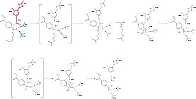
In a second step, a water molecule is deprotonated by H242 and D210, allowing it to nucleophilically attack the carbonyl carbon, forming a tetrahedral intermediate and an oxyanion that is stabilized by the same <<scene name='10/1075193/Oxyanion_hole/2'>oxyanion hole</scene>. H242 protonates the leaving group oxygen of S165, allowing the reformation of the carbonyl and the severing of the covalent bond to serine. The -1 monomer is released from the enzyme, and the protons are reset for further catalysis. | In a second step, a water molecule is deprotonated by H242 and D210, allowing it to nucleophilically attack the carbonyl carbon, forming a tetrahedral intermediate and an oxyanion that is stabilized by the same <<scene name='10/1075193/Oxyanion_hole/2'>oxyanion hole</scene>. H242 protonates the leaving group oxygen of S165, allowing the reformation of the carbonyl and the severing of the covalent bond to serine. The -1 monomer is released from the enzyme, and the protons are reset for further catalysis. | ||
[[Image:PET_hydrolase_Mechanism.jpeg|400 px|left|thumb|Figure Legend]] <b>need higher quality image of this</b> | [[Image:PET_hydrolase_Mechanism.jpeg|400 px|left|thumb|Figure Legend]] <b>need higher quality image of this</b> | ||
| - | =Mutations= | + | ==Mutations== |
Researchers have been investigating various mutations of PET hydrolase to enhance its catalytic ability. One group of researchers, Tournier et. al., have made mutations in the PET hydrolase active site. They identified the key residues involved in the catalytic mechanism by using a model of the <scene name='10/1075190/Ligand/2'>PET substrate</scene> onto the enzyme. The site, mainly a hydrophobic pocket, contained 11 residues targeted for mutagenesis. From this, they identified that the majority of enzymes' specific activity went down; however, the mutation of the F243 to either isoleucine or tryptophan increased specific activity. Four target mutations introduced into the PET Hydrolase by Tournier et. al. demonstrated improved catalytic efficiency and thermal stability compared to its wild-type structure. | Researchers have been investigating various mutations of PET hydrolase to enhance its catalytic ability. One group of researchers, Tournier et. al., have made mutations in the PET hydrolase active site. They identified the key residues involved in the catalytic mechanism by using a model of the <scene name='10/1075190/Ligand/2'>PET substrate</scene> onto the enzyme. The site, mainly a hydrophobic pocket, contained 11 residues targeted for mutagenesis. From this, they identified that the majority of enzymes' specific activity went down; however, the mutation of the F243 to either isoleucine or tryptophan increased specific activity. Four target mutations introduced into the PET Hydrolase by Tournier et. al. demonstrated improved catalytic efficiency and thermal stability compared to its wild-type structure. | ||
==F243I/W Mutations== | ==F243I/W Mutations== | ||
| Line 65: | Line 65: | ||
==Glycosylation== | ==Glycosylation== | ||
<scene name='10/1075191/All_glycosylation_sites/5'>Glycosylation sites</scene> were introduced in a research study completed by Abhihit N. Shirke and others with the initial intention of inducing aggregation in the leaf branch cutinase / PET hydrolase wild-type. <ref name="Shirke">PMID:29328676</ref> Glycosylation, as a general tool, is introduced into a protein to improve conformational stability. The specific type used in this study was N-linked side chain alteration. This means that the glycosylation sites were selected based on a starting asparagine residue followed by the sequence N-X-S or N-X-T, where X stands for any of the twenty amino acids except proline. <ref name="Imperiali">PMID:10600722</ref> This is implemented specifically because it allows for a better ability to choose the mutation sites. The first glycosylation site followed the N-T-S pattern with <scene name='10/1075191/First_glycosylation_site/5'>residues 197-199</scene>. The second was <scene name='10/1075191/Second_glycosylation_site/4'>residues 239-241</scene> with an N-A-S pattern, located nearest to the active site. The final was <scene name='10/1075191/Third_glycosylation_site/4'>residues 266-268</scene>, exhibiting a N-D-T sequence. <ref name="Shirke">PMID:29328676</ref> With the combination of these glycosylation sites (without any other mutagenesis introduced to the enzyme), an overall 10-degree Celsius higher thermal stability was exhibited compared to the wild-type, with the structure of the modified PET hydrolase three times more stable. Catalytic efficiency also improved at the enzyme's known melting temperature. Even though the target point of introducing glycosylation sites was to induce aggregation in the PET hydrolase, depletion of aggregation was exhibited. The glycosylated protein took twice as long to unfold compared to the wild-type. For comparison, the threshold of 80 degrees Celsius was where the major difference in kinetic activity occurred: 85% of the glycosylated hydrolase maintained its catalytic activity, whereas the wild-type only had 50% of it working at the same temperature.<ref name="Shirke">PMID:29328676</ref> The first and third sites showed these trends both together and on their own as the only glycosylation sites, but the second one nearest the active site showed no change when glycosylated on its own when compared to the wild type. <ref name="Shirke">PMID:29328676</ref> | <scene name='10/1075191/All_glycosylation_sites/5'>Glycosylation sites</scene> were introduced in a research study completed by Abhihit N. Shirke and others with the initial intention of inducing aggregation in the leaf branch cutinase / PET hydrolase wild-type. <ref name="Shirke">PMID:29328676</ref> Glycosylation, as a general tool, is introduced into a protein to improve conformational stability. The specific type used in this study was N-linked side chain alteration. This means that the glycosylation sites were selected based on a starting asparagine residue followed by the sequence N-X-S or N-X-T, where X stands for any of the twenty amino acids except proline. <ref name="Imperiali">PMID:10600722</ref> This is implemented specifically because it allows for a better ability to choose the mutation sites. The first glycosylation site followed the N-T-S pattern with <scene name='10/1075191/First_glycosylation_site/5'>residues 197-199</scene>. The second was <scene name='10/1075191/Second_glycosylation_site/4'>residues 239-241</scene> with an N-A-S pattern, located nearest to the active site. The final was <scene name='10/1075191/Third_glycosylation_site/4'>residues 266-268</scene>, exhibiting a N-D-T sequence. <ref name="Shirke">PMID:29328676</ref> With the combination of these glycosylation sites (without any other mutagenesis introduced to the enzyme), an overall 10-degree Celsius higher thermal stability was exhibited compared to the wild-type, with the structure of the modified PET hydrolase three times more stable. Catalytic efficiency also improved at the enzyme's known melting temperature. Even though the target point of introducing glycosylation sites was to induce aggregation in the PET hydrolase, depletion of aggregation was exhibited. The glycosylated protein took twice as long to unfold compared to the wild-type. For comparison, the threshold of 80 degrees Celsius was where the major difference in kinetic activity occurred: 85% of the glycosylated hydrolase maintained its catalytic activity, whereas the wild-type only had 50% of it working at the same temperature.<ref name="Shirke">PMID:29328676</ref> The first and third sites showed these trends both together and on their own as the only glycosylation sites, but the second one nearest the active site showed no change when glycosylated on its own when compared to the wild type. <ref name="Shirke">PMID:29328676</ref> | ||
| - | =Biochemical Results= | + | ==Biochemical Results== |
==Improved Thermal Stability== | ==Improved Thermal Stability== | ||
Thermal stability is very important for enzyme-catalyzed PET degradation because the reaction must take place above the transition temperature of PET(70ºC), which allows the substrate to have optimal flexibility to fit into the active site. The disulfide bridge mutation raises the melting point of the enzyme from 84.7ºC to 94.5ºC. | Thermal stability is very important for enzyme-catalyzed PET degradation because the reaction must take place above the transition temperature of PET(70ºC), which allows the substrate to have optimal flexibility to fit into the active site. The disulfide bridge mutation raises the melting point of the enzyme from 84.7ºC to 94.5ºC. | ||
| Line 72: | Line 72: | ||
==Enhanced Catalytic Efficiency== | ==Enhanced Catalytic Efficiency== | ||
The ICCG and WCCG mutations constructed by Tournier et al. showed a return to the wild type activity and beyond. The tryptophan mutation showed a 122% increase in catalysis, with an increased ability to sustain its structure at temperatures 6.2 degrees higher than the wild type (84.7 degrees Celsius). The isoleucine mutation showed a 2% decrease in activity compared to the wild type, but a thermal stability increase by 10.1 degrees Celsius. | The ICCG and WCCG mutations constructed by Tournier et al. showed a return to the wild type activity and beyond. The tryptophan mutation showed a 122% increase in catalysis, with an increased ability to sustain its structure at temperatures 6.2 degrees higher than the wild type (84.7 degrees Celsius). The isoleucine mutation showed a 2% decrease in activity compared to the wild type, but a thermal stability increase by 10.1 degrees Celsius. | ||
| - | =Conclusions= | + | ==Conclusions== |
==Implications for Plastic Recycling== | ==Implications for Plastic Recycling== | ||
Information here | Information here | ||
Revision as of 14:13, 10 April 2025
PET hydrolase
| |||||||||||
Student Contributors
- Georgia Apple
- Emily Hwang
- Anjali Rabindran

