We apologize for Proteopedia being slow to respond. For the past two years, a new implementation of Proteopedia has been being built. Soon, it will replace this 18-year old system. All existing content will be moved to the new system at a date that will be announced here.
User:Carson Powers/Sandbox 1
From Proteopedia
(Difference between revisions)
| Line 6: | Line 6: | ||
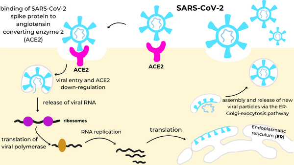
[[Image:Infection_Cycle.png|600px|right|SARS-CoV-2 infection cycle: the viral spike protein (cyan) binds to the ACE2 receptor (magenta) on the cell surface, triggering endocytosis and ACE2 down-regulation, release of viral RNA into the cytosol, translation of the viral polymerase and genome replication, synthesis of structural proteins in the endoplasmic reticulum (ER), and assembly and release of new virions via the ER-Golgi-exocytosis pathway.]] | [[Image:Infection_Cycle.png|600px|right|SARS-CoV-2 infection cycle: the viral spike protein (cyan) binds to the ACE2 receptor (magenta) on the cell surface, triggering endocytosis and ACE2 down-regulation, release of viral RNA into the cytosol, translation of the viral polymerase and genome replication, synthesis of structural proteins in the endoplasmic reticulum (ER), and assembly and release of new virions via the ER-Golgi-exocytosis pathway.]] | ||
| - | |||
=Spike Protein= | =Spike Protein= | ||
| - | |||
To begin, the spike protein[https://en.wikipedia.org/wiki/Coronavirus_spike_protein] is a homotrimer glycoprotein that lies on the surface of SARS-CoV-2 to facilitate the entry of the virus into host cells. The spike utilizes the Angiotensin Converting Enzyme Receptor 2 (ACE2)[https://en.wikipedia.org/wiki/Angiotensin-converting_enzyme_2] in the lungs to enter. Once inside the cell, virus-specific RNA and proteins are synthesized within the cytoplasm. The viral envelope merges with the oily membrane of our own cells, allowing the virus to release its genetic material into the inside of the healthy cell. Each infected cell may produce and release millions of copies of the virus which can infect other neighboring cells and people when the viral particles are released from the airways (i.e., via coughing or sneezing). This pathway is visualized in the image to the right. | To begin, the spike protein[https://en.wikipedia.org/wiki/Coronavirus_spike_protein] is a homotrimer glycoprotein that lies on the surface of SARS-CoV-2 to facilitate the entry of the virus into host cells. The spike utilizes the Angiotensin Converting Enzyme Receptor 2 (ACE2)[https://en.wikipedia.org/wiki/Angiotensin-converting_enzyme_2] in the lungs to enter. Once inside the cell, virus-specific RNA and proteins are synthesized within the cytoplasm. The viral envelope merges with the oily membrane of our own cells, allowing the virus to release its genetic material into the inside of the healthy cell. Each infected cell may produce and release millions of copies of the virus which can infect other neighboring cells and people when the viral particles are released from the airways (i.e., via coughing or sneezing). This pathway is visualized in the image to the right. | ||
| Line 26: | Line 24: | ||
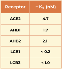
Another miniprotein, <scene name='10/1076041/Lcb3_scene_1/4'>LCB3</scene>, was developed using the same computational method as LCB1 and binds to the same region of the RBD, except in the opposite direction. Its <scene name='10/1076041/Lcb3xspikecorlabel_intxn/1'>binding interactions</scene> also show a large number of hydrogen bonds. However, LCB1 has more surface area and fits more precisely with the spike protein, giving it a slightly higher binding affinity (shown in the table to the right).[[Image:Kd_table.png|200px|right|'''Table 1'''. Dissociation constants (Kd) in nM for SARS-CoV-2 spike RBD binding to ACE2 and engineered minibinders, measured by biolayer interferometry to compare affinities for the spike protein.]] | Another miniprotein, <scene name='10/1076041/Lcb3_scene_1/4'>LCB3</scene>, was developed using the same computational method as LCB1 and binds to the same region of the RBD, except in the opposite direction. Its <scene name='10/1076041/Lcb3xspikecorlabel_intxn/1'>binding interactions</scene> also show a large number of hydrogen bonds. However, LCB1 has more surface area and fits more precisely with the spike protein, giving it a slightly higher binding affinity (shown in the table to the right).[[Image:Kd_table.png|200px|right|'''Table 1'''. Dissociation constants (Kd) in nM for SARS-CoV-2 spike RBD binding to ACE2 and engineered minibinders, measured by biolayer interferometry to compare affinities for the spike protein.]] | ||
| - | |||
=Significance= | =Significance= | ||
Revision as of 18:18, 28 April 2025
De Novo Miniprotein COVID-19 Therapeutic
| |||||||||||


