User:Hayden Vissing/Sandbox 1
From Proteopedia
< User:Hayden Vissing(Difference between revisions)
| Line 28: | Line 28: | ||
==Mutations== | ==Mutations== | ||
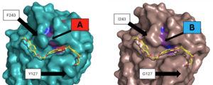
| - | After selection, LCC was mutated in order to improve its efficiency and thermal stability. Tournier et al. (2020)<ref name="main" /> identified <scene name='10/1075218/Wildtype_residues/5'>11 residues</scene> from the wildtype as being apart of the <scene name='10/1076051/Contact_shell/2'>first contact shell</scene>. They then made hundreds of different mutation | + | After selection, LCC was mutated in order to improve its efficiency and thermal stability. Tournier et al. (2020)<ref name="main" /> identified <scene name='10/1075218/Wildtype_residues/5'>11 residues</scene> from the wildtype as being apart of the <scene name='10/1076051/Contact_shell/2'>first contact shell</scene>. They then made hundreds of different mutation combinations, marking which altered efficiency and stability the most. Two of these 11 were chosen for the final mutant enzyme, <scene name='10/1075218/Identifiedresidues/13'>F243I and Y127G</scene>. These mutations were paired with two separately identified thermostability specific mutations (D238C and S283C) to make the final <scene name='10/1076051/Mutant_overview_shade/3'>ICCG</scene> enzyme (PDB: 6THT<ref name="6tht">Sulaiman S, You DJ, Eiko K, Koga Y, Kanaya S. High resolution crystal structure of a leaf-branch compost cutinase homolog. RCSB Protein Data Bank. Published December 4, 2019. https://www.rcsb.org/structure/6THT</ref>). |
===Efficiency improvement=== | ===Efficiency improvement=== | ||
| Line 37: | Line 37: | ||
===Thermostability improvement=== | ===Thermostability improvement=== | ||
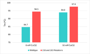
[[Image:Temperatures.png|300px|right|thumb|Figure 5 - Thermostability lab data from Tournier et al. (2020)<ref name="main" />]] | [[Image:Temperatures.png|300px|right|thumb|Figure 5 - Thermostability lab data from Tournier et al. (2020)<ref name="main" />]] | ||
| - | To allow the enzyme to be effective in the recycling of PET, it needed to be stable in the high temperature environments used during the recycling process. Because of this, improvements needed to be made to the thermostability of the enzyme. The researchers went about this by searching for divalent metal binding sites, which had been seen to improve the thermal stability of other PET hydrolase enzymes<ref name="main" />. They found this site formed by the residues of <scene name='10/1076051/3res_before/2'>E208, D238, and S283</scene>. These residues were picked from LCC due to being in the same location of a metal binding site that is found on <scene name='10/1076051/3_res/2'>other known cutinases</scene>. All 3 including LCC shared the same 2 to 3 acidic residues which allow a calcium or magnesium ion to bind. that had been seen in other PET hydrolase enzymes, and when calcium ions were added to the enzyme the thermal stability improved. In order to avoid issues with purification, the team opted to avoid the addition of calcium salts and to instead create a disulfide bridge at the location of the divalent metal binding site. They chose to replace the <scene name='10/1076054/Thermostability_wild_type/5'>aspartic acid at 238 and the serine at 283</scene> with cysteine residues to form this <scene name='10/1076054/Iccg_disulfide_bridge/6'>disulfide bridge</scene>. This mutation improved the melting temperature of the enzyme by 9.8℃<ref name="main" />. Figure 5 uses this thermostability data and displays visually the effects the mutations have on melting temperatures<ref name="main" />. | + | To allow the enzyme to be effective in the recycling of PET, it needed to be stable in the high temperature environments used during the recycling process. Because of this, improvements needed to be made to the thermostability of the enzyme. The researchers went about this by searching for divalent metal binding sites, which had been seen to improve the thermal stability of other PET hydrolase enzymes<ref name="main" />. They found this site formed by the residues of <scene name='10/1076051/3res_before/2'>E208, D238, and S283</scene>. These residues were picked from LCC due to being in the same location of a metal binding site that is found on <scene name='10/1076051/3_res/2'>other known cutinases</scene>. All 3 including LCC shared the same 2 to 3 acidic residue motif that allowed a metal ion to bind. When calcium ions were added, the thermal stability improved. (reference). All 3 including LCC shared the same 2 to 3 acidic residues which allow a calcium or magnesium ion to bind. that had been seen in other PET hydrolase enzymes, and when calcium ions were added to the enzyme the thermal stability improved. In order to avoid issues with purification, the team opted to avoid the addition of calcium salts and to instead create a disulfide bridge at the location of the divalent metal binding site. They chose to replace the <scene name='10/1076054/Thermostability_wild_type/5'>aspartic acid at 238 and the serine at 283</scene> with cysteine residues to form this <scene name='10/1076054/Iccg_disulfide_bridge/6'>disulfide bridge</scene>. This mutation improved the melting temperature of the enzyme by 9.8℃<ref name="main" />. Figure 5 uses this thermostability data and displays visually the effects the mutations have on melting temperatures<ref name="main" />. |
==Conclusions== | ==Conclusions== | ||
Current revision
The Future of Recycling: PET Hydrolase Enzyme with Improved Efficiency and Stability
| |||||||||||
References
- ↑ 1.0 1.1 Hiraga, K., Taniguchi, I., Yoshida, S., Kimura, Y., & Oda, K. (2019). Biodegradation of waste PET: A sustainable solution for dealing with plastic pollution. EMBO Reports, 20(11), e49365. https://doi.org/10.15252/embr.201949365. [Published correction appears in EMBO Reports, 21(2), e49826. DOI: 10.15252/embr.201949826
- ↑ 2.0 2.1 2.2 2.3 Jayasekara, S. K., Joni, H. D., Jayantha, B., Dissanayake, L., Mandrell, C., Sinharage, M. M. S., Molitor, R., Jayasekara, T., Sivakumar, P., & Jayakody, L. N. (2023). Trends in in-silico guided engineering of efficient polyethylene terephthalate (PET) hydrolyzing enzymes to enable bio-recycling and upcycling of PET. Computational and structural biotechnology journal, 21, 3513–3521. DOI: 10.1016/j.csbj.2023.06.004
- ↑ 3.0 3.1 Babaei, M., Jalilian, M., & Shahbaz, K. (2024). Chemical recycling of Polyethylene terephthalate: A mini-review. Journal of Environmental Chemical Engineering, 12(3), 112507. DOI: 10.1016/j.jece.2024.112507
- ↑ 4.00 4.01 4.02 4.03 4.04 4.05 4.06 4.07 4.08 4.09 4.10 4.11 Tournier V, Topham CM, Gilles A, David B, Folgoas C, Moya-Leclair E, Kamionka E, Desrousseaux ML, Texier H, Gavalda S, Cot M, Guemard E, Dalibey M, Nomme J, Cioci G, Barbe S, Chateau M, Andre I, Duquesne S, Marty A. An engineered PET depolymerase to break down and recycle plastic bottles. Nature. 2020 Apr;580(7802):216-219. doi: 10.1038/s41586-020-2149-4. Epub 2020 Apr, 8. PMID:32269349 doi:http://dx.doi.org/10.1038/s41586-020-2149-4
- ↑ Sulaiman S, You DJ, Eiko K, Koga Y, Kanaya S. Crystal structure of leaf-branch compost bacterial cutinase homolog. RCSB Protein Data Bank. Published March 27, 2013. https://www.rcsb.org/structure/4EB0
- ↑ Joo S, Cho IJ, Seo H, Son HF, Sagong HY, Shin TJ, Choi SY, Lee SY, Kim KJ. Structural insight into molecular mechanism of poly(ethylene terephthalate) degradation. Nat Commun. 2018 Jan 26;9(1):382. doi: 10.1038/s41467-018-02881-1. PMID:29374183 doi:http://dx.doi.org/10.1038/s41467-018-02881-1
- ↑ Sulaiman S, Yamato S, Kanaya E, Kim JJ, Koga Y, Takano K, Kanaya S. Isolation of a novel cutinase homolog with polyethylene terephthalate-degrading activity from leaf-branch compost by using a metagenomic approach. Appl Environ Microbiol. 2012 Mar;78(5):1556-62. doi: 10.1128/AEM.06725-11. Epub, 2011 Dec 22. PMID:22194294 doi:http://dx.doi.org/10.1128/AEM.06725-11
- ↑ Boneta S, Arafet K, Moliner V. QM/MM Study of the Enzymatic Biodegradation Mechanism of Polyethylene Terephthalate. J Chem Inf Model. 2021;61(6):3041-3051. doi:10.1021/acs.jcim.1c00394
- ↑ A binding model of the substrate 2-HE(MHET)3 in wild-type LLC (4eb0.pdb) was constructed and refined to mimic the 3D structure illustrated in Figure 2 of reference “3”. The software Maestro (Schrödinger, Inc; version 14.2.118) was used to construct the initial binding structure, followed by energy minimization in the context of the rigid protein that had previously been processed to add/refine all hydrogen atoms. The ligand model was then used without further modification to identify and illustrate the cited active-site residues.
- ↑ Han, X., Liu, W., Huang, J. W., et al. (2017). Structural insight into catalytic mechanism of PET hydrolase. Nature Communications, 8, 2106. https://doi.org/10.1038/s41467-017-02255-z.Heredia-Guerrero, J. A., Heredia, A., García-Segura, R., & Benítez, J. J. (2009). Synthesis and characterization of a plant cutin mimetic polymer. Polymer, 50(24), 5633–5637. DOI: 10.1016/j.polymer.2009.10.018
- ↑ Sulaiman S, You DJ, Eiko K, Koga Y, Kanaya S. High resolution crystal structure of a leaf-branch compost cutinase homolog. RCSB Protein Data Bank. Published December 4, 2019. https://www.rcsb.org/structure/6THT
Additional Literature and Resources
- Roth C, Wei R, Oeser T, Then J, Follner C, Zimmermann W, Strater N. Structural and functional studies on a thermostable polyethylene terephthalate degrading hydrolase from Thermobifida fusca. Appl Microbiol Biotechnol. 2014 Apr 13. PMID:24728714 doi:http://dx.doi.org/10.1007/s00253-014-5672-0
Student Contributors
- David Bogle
- Justin Chavez
- Hayden Vissing
