We apologize for Proteopedia being slow to respond. For the past two years, a new implementation of Proteopedia has been being built. Soon, it will replace this 18-year old system. All existing content will be moved to the new system at a date that will be announced here.
User:Karsten Theis/T7RNAP physical model explanation
From Proteopedia
(Difference between revisions)
| Line 6: | Line 6: | ||
[[Image:1msw legend.png|800px]] | [[Image:1msw legend.png|800px]] | ||
| - | <StructureSection load='' size='340' side='right' caption='' scene='10/ | + | <StructureSection load='' size='340' side='right' caption='' scene='10/1081719/T7_rnap/1'> |
<html5media height="400" width="300">https://www.youtube.com/watch?v=q3GUgPdEBbU</html5media> | <html5media height="400" width="300">https://www.youtube.com/watch?v=q3GUgPdEBbU</html5media> | ||
Revision as of 02:37, 20 June 2025
This is a tour of a physical model of the T7 virus RNA polymerase (PDB ID 1msw).

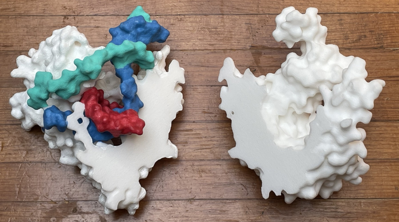
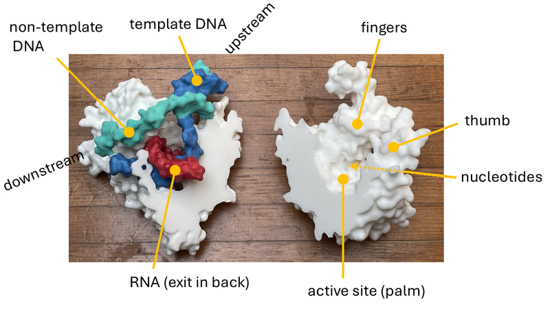
Above is a photograph of the two parts of the model. To assemble it, you bring the two flat sides together (the view is a "butterflied" version of the structure). On the left is the N-terminal part (roughly) with the nucleic acid forming a transcription bubble. On the right is the C-terminal part (roughly), in the shape of the right hand, with the active site at the palm. For an annotated image, see below.
| |||||||||||

