Sandbox Home
From Proteopedia
| Line 24: | Line 24: | ||
</tr> | </tr> | ||
| - | <!-- SINGLE CONTENT ROW | + | <!-- SINGLE CONTENT ROW --> |
| - | <tr | + | <tr> |
| - | <!-- LEFT COLUMN | + | <!-- LEFT COLUMN --> |
| - | <td style="padding:10px;"> | + | <td style="padding:10px; vertical-align:top !important;"> |
| - | <!-- Links ( | + | <!-- Links first (will sit at top) --> |
<div> | <div> | ||
<p>[[Help:Contents#For_authors:_contributing_content|How to add content to Proteopedia]]</p> | <p>[[Help:Contents#For_authors:_contributing_content|How to add content to Proteopedia]]</p> | ||
| Line 35: | Line 35: | ||
</div> | </div> | ||
| - | <!-- Featured | + | <!-- Featured underneath --> |
<div style="margin-top:10px;"> | <div style="margin-top:10px;"> | ||
{{Proteopedia:Featured SEL/{{#expr: {{#time:U}} mod {{Proteopedia:Number of SEL articles}}}}}} | {{Proteopedia:Featured SEL/{{#expr: {{#time:U}} mod {{Proteopedia:Number of SEL articles}}}}}} | ||
| Line 42: | Line 42: | ||
<!-- MIDDLE COLUMN --> | <!-- MIDDLE COLUMN --> | ||
| - | <td style="padding:10px;"> | + | <td style="padding:10px; vertical-align:top !important;"> |
<div> | <div> | ||
<p>[[I3DC|About Interactive 3D Complements - '''I3DCs''']]</p> | <p>[[I3DC|About Interactive 3D Complements - '''I3DCs''']]</p> | ||
| Line 48: | Line 48: | ||
<p>[[How to get an I3DC for your paper]]</p> | <p>[[How to get an I3DC for your paper]]</p> | ||
</div> | </div> | ||
| - | |||
<div style="margin-top:10px;"> | <div style="margin-top:10px;"> | ||
{{Proteopedia:Featured JRN/{{#expr: {{#time:U}} mod {{Proteopedia:Number of JRN articles}}}}}} | {{Proteopedia:Featured JRN/{{#expr: {{#time:U}} mod {{Proteopedia:Number of JRN articles}}}}}} | ||
| Line 55: | Line 54: | ||
<!-- RIGHT COLUMN --> | <!-- RIGHT COLUMN --> | ||
| - | <td style="padding:10px;"> | + | <td style="padding:10px; vertical-align:top !important;"> |
<div> | <div> | ||
<p>[[Teaching strategies using Proteopedia]]</p> | <p>[[Teaching strategies using Proteopedia]]</p> | ||
| Line 61: | Line 60: | ||
<p>[[Help:Contents#For_authors:_contributing_content|How to add content to Proteopedia]]</p> | <p>[[Help:Contents#For_authors:_contributing_content|How to add content to Proteopedia]]</p> | ||
</div> | </div> | ||
| - | |||
<div style="margin-top:10px;"> | <div style="margin-top:10px;"> | ||
{{Proteopedia:Featured EDU/{{#expr: {{#time:U}} mod {{Proteopedia:Number of EDU articles}}}}}} | {{Proteopedia:Featured EDU/{{#expr: {{#time:U}} mod {{Proteopedia:Number of EDU articles}}}}}} | ||
Revision as of 16:00, 30 September 2025
|
ISSN 2310-6301
As life is more than 2D, Proteopedia helps to bridge the gap between 3D structure & function of biomacromolecules
Proteopedia presents this information in a user-friendly way as a collaborative & free 3D-encyclopedia of proteins & other biomolecules.
|
||||||||
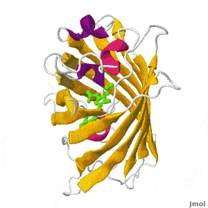
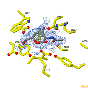
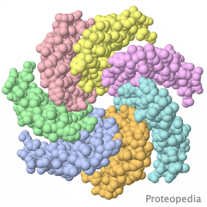
| Selected Research Pages | In Journals | Education | ||||||
|---|---|---|---|---|---|---|---|---|
|
|
|
||||||
|
||||||||