This old version of Proteopedia is provided for student assignments while the new version is undergoing repairs. Content and edits done in this old version of Proteopedia after March 1, 2026 will eventually be lost when it is retired in about June of 2026.
Apply for new accounts at the new Proteopedia. Your logins will work in both the old and new versions.
1x9d
From Proteopedia
| Line 1: | Line 1: | ||
[[Image:1x9d.gif|left|200px]] | [[Image:1x9d.gif|left|200px]] | ||
| - | + | <!-- | |
| - | + | The line below this paragraph, containing "STRUCTURE_1x9d", creates the "Structure Box" on the page. | |
| - | + | You may change the PDB parameter (which sets the PDB file loaded into the applet) | |
| - | + | or the SCENE parameter (which sets the initial scene displayed when the page is loaded), | |
| - | + | or leave the SCENE parameter empty for the default display. | |
| - | | | + | --> |
| - | | | + | {{STRUCTURE_1x9d| PDB=1x9d | SCENE= }} |
| - | + | ||
| - | + | ||
| - | }} | + | |
'''Crystal Structure Of Human Class I alpha-1,2-Mannosidase In Complex With Thio-Disaccharide Substrate Analogue''' | '''Crystal Structure Of Human Class I alpha-1,2-Mannosidase In Complex With Thio-Disaccharide Substrate Analogue''' | ||
| Line 32: | Line 29: | ||
[[Category: Tempel, W.]] | [[Category: Tempel, W.]] | ||
[[Category: Wang, B C.]] | [[Category: Wang, B C.]] | ||
| - | [[Category: | + | [[Category: Glycosyl hydrolase]] |
| - | [[Category: | + | [[Category: Mannosidase]] |
| - | [[Category: | + | [[Category: Substrate analogue]] |
| - | + | ''Page seeded by [http://oca.weizmann.ac.il/oca OCA ] on Sat May 3 14:44:13 2008'' | |
| - | ''Page seeded by [http://oca.weizmann.ac.il/oca OCA ] on | + | |
Revision as of 11:44, 3 May 2008
| |||||||||
| 1x9d, resolution 1.41Å () | |||||||||
|---|---|---|---|---|---|---|---|---|---|
| Ligands: | , , , | ||||||||
| Gene: | MAN1B1 (Homo sapiens) | ||||||||
| Activity: | Mannosyl-oligosaccharide 1,2-alpha-mannosidase, with EC number 3.2.1.113 | ||||||||
| Related: | 1fmi | ||||||||
| |||||||||
| |||||||||
| Resources: | FirstGlance, OCA, RCSB, PDBsum | ||||||||
| Coordinates: | save as pdb, mmCIF, xml | ||||||||
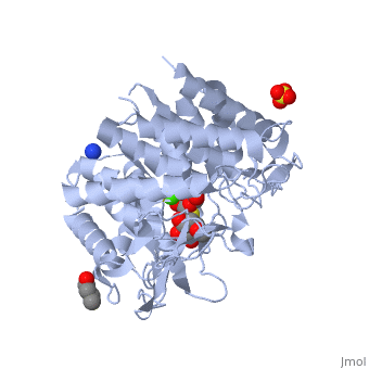

Crystal Structure Of Human Class I alpha-1,2-Mannosidase In Complex With Thio-Disaccharide Substrate Analogue
Overview
Quality control in the endoplasmic reticulum (ER) determines the fate of newly synthesized glycoproteins toward either correct folding or disposal by ER-associated degradation. Initiation of the disposal process involves selective trimming of N-glycans attached to misfolded glycoproteins by ER alpha-mannosidase I and subsequent recognition by the ER degradation-enhancing alpha-mannosidase-like protein family of lectins, both members of glycosylhydrolase family 47. The unusual inverting hydrolytic mechanism catalyzed by members of this family is investigated here by a combination of kinetic and binding analyses of wild type and mutant forms of human ER alpha-mannosidase I as well as by structural analysis of a co-complex with an uncleaved thiodisaccharide substrate analog. These data reveal the roles of potential catalytic acid and base residues and the identification of a novel (3)S(1) sugar conformation for the bound substrate analog. The co-crystal structure described here, in combination with the (1)C(4) conformation of a previously identified co-complex with the glycone mimic, 1-deoxymannojirimycin, indicates that glycoside bond cleavage proceeds through a least motion conformational twist of a properly predisposed substrate in the -1 subsite. A novel (3)H(4) conformation is proposed as the exploded transition state.
About this Structure
1X9D is a Single protein structure of sequence from Homo sapiens. Full crystallographic information is available from OCA.
Reference
Mechanism of class 1 (glycosylhydrolase family 47) {alpha}-mannosidases involved in N-glycan processing and endoplasmic reticulum quality control., Karaveg K, Siriwardena A, Tempel W, Liu ZJ, Glushka J, Wang BC, Moremen KW, J Biol Chem. 2005 Apr 22;280(16):16197-207. Epub 2005 Feb 15. PMID:15713668 Page seeded by OCA on Sat May 3 14:44:13 2008
