This old version of Proteopedia is provided for student assignments while the new version is undergoing repairs. Content and edits done in this old version of Proteopedia after March 1, 2026 will eventually be lost when it is retired in about June of 2026.
Apply for new accounts at the new Proteopedia. Your logins will work in both the old and new versions.
Temperature value
From Proteopedia
(→Coloring by Temperature - adding content) |
(→Coloring by Temperature - adding content) |
||
| Line 11: | Line 11: | ||
Visualizing relative disorder or uncertainty in atomic positions is done by ''coloring by temperature value''. Atoms with <font color='blue'>'''low temperature values are colored blue'''</font>, while atoms with <font color='red'>'''high temperature values are colored red'''</font>. Light blue, white, and pink atoms represent the scale of intermediate temperature values. The temperature values themselves are relative, not absolute, and hence the colors are also relative. The most important information from coloring by temperature is to identify the ''red'' residues whose positions are least certain. Their positions should be taken only as rough approximations, and this is especially worth knowing if any residues in areas of special interest are red. | Visualizing relative disorder or uncertainty in atomic positions is done by ''coloring by temperature value''. Atoms with <font color='blue'>'''low temperature values are colored blue'''</font>, while atoms with <font color='red'>'''high temperature values are colored red'''</font>. Light blue, white, and pink atoms represent the scale of intermediate temperature values. The temperature values themselves are relative, not absolute, and hence the colors are also relative. The most important information from coloring by temperature is to identify the ''red'' residues whose positions are least certain. Their positions should be taken only as rough approximations, and this is especially worth knowing if any residues in areas of special interest are red. | ||
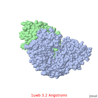
| - | While the temperature colors of two separate models cannot be rigorously compared (since the temperature values themselves are relative, not absolute), high [[Resolution|resolution]] models tend to have fewer red atoms than do models with modest resolution. For example, a <scene name='Temperature_value/1kzk/1'>small HIV protease at 1.1 Angstroms resolution</scene> ([[1kzk]]) has few red atoms, while the <scene name='Temperature_value/1uwb/2'>larger, less compact HIV reverse transcriptase at 3.2 Angstroms resolution</scene> ([[1uwb]]) has <scene name='Temperature_value/1uwb/1'>many red atoms</scene>. Also, many of the surface sidechains are missing (due to disorder) in [[1uwb]]. Missing sidechains are labeled '''S-''' in ''FirstGlance in Jmol'' (linked under the molecule on all [[PDB code]]-titled pages in Proteopedia). | + | While the temperature colors of two separate models cannot be rigorously compared (since the temperature values themselves are relative, not absolute), high [[Resolution|resolution]] models tend to have fewer red atoms than do models with modest resolution. For example, a <scene name='Temperature_value/1kzk/1'>small HIV protease at 1.1 Angstroms resolution</scene> ([[1kzk]]) has <scene name='Temperature_value/1kzk/2'>few red atoms</scene>, while the <scene name='Temperature_value/1uwb/2'>larger, less compact HIV reverse transcriptase at 3.2 Angstroms resolution</scene> ([[1uwb]]) has <scene name='Temperature_value/1uwb/1'>many red atoms</scene>. Also, many of the surface sidechains are missing (due to disorder) in [[1uwb]]. Missing sidechains are labeled '''S-''' in ''FirstGlance in Jmol'' (linked under the molecule on all [[PDB code]]-titled pages in Proteopedia). |
==Missing Residues== | ==Missing Residues== | ||
Revision as of 01:24, 27 June 2008
Contents |
Definition
In crystallography, uncertainty in the positions of atoms increases with disorder in the protein crystal. Disorder may have two components, static and dynamic. First, some regions of the molecule may adopt different conformations in different copies of the molecule, each molecule's conformation being stable (static disorder). Second, some regions of every copy of the molecule may be subject to thermal motion, meaning vibration about the rest position[1] Thermal motion is minimized when the crystal is frozen with liquid nitrogen while being irradiated, but irradiation may warm the crystal permitting some thermal motion.
Some regions of the molecule may have higher average disorder, and others lower average disorder. Typically, the ends of chains have higher average disorder, and hence their positions are less certain than are residues in the core of a tightly packed domain, where disorder is less. The disorder for each atom is quantitated in its temperature value.
Coloring by Temperature
|
Visualizing relative disorder or uncertainty in atomic positions is done by coloring by temperature value. Atoms with low temperature values are colored blue, while atoms with high temperature values are colored red. Light blue, white, and pink atoms represent the scale of intermediate temperature values. The temperature values themselves are relative, not absolute, and hence the colors are also relative. The most important information from coloring by temperature is to identify the red residues whose positions are least certain. Their positions should be taken only as rough approximations, and this is especially worth knowing if any residues in areas of special interest are red.
While the temperature colors of two separate models cannot be rigorously compared (since the temperature values themselves are relative, not absolute), high resolution models tend to have fewer red atoms than do models with modest resolution. For example, a (1kzk) has , while the (1uwb) has . Also, many of the surface sidechains are missing (due to disorder) in 1uwb. Missing sidechains are labeled S- in FirstGlance in Jmol (linked under the molecule on all PDB code-titled pages in Proteopedia).
Missing Residues
Often the very ends of chains, or surface loops, may be so disordered as to prevent assigning any atomic positions at all, leading to missing residues. FirstGlance in Jmol (linked beneath the molecule on every PDB code-titled page in Proteopedia) has a Gaps button that explains how to detect and visualize missing residues.
Data Format
In the PDB file format, each atom is given not only X, Y, and Z Cartesian coordinates, but two additional values immediately following called occupancy and temperature value (also known as the isotropic B value). If the end of a chain adopts either of two stable positions with equal probability, each position has 50% occupancy. The temperature factor is provided to quantitate the level of thermal motion. However, these two components of disorder cannot be distinguished with crystal diffraction data alone. Therefore, the occupancy is often given as 1.0 (100%), while the degree of "blur" in the electron density map, representing both components of disorder, is reported in the temperature value field. These values are mapped to colors when a crystallographic result is colored by temperature.
Uncertainty in NMR Models
NMR models provide no information in the temperature value fields of PDB files. Rather, the variation between the models gives some indication of uncertainty or flexibility.
See Also
Content Donors
Large portions of this page were adapted from the Glossary of ProteinExplorer.Org, with the permission of the principal author, Eric Martz.
