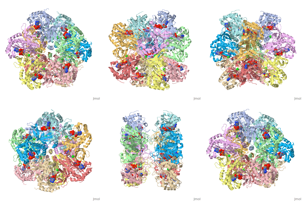
User:Tom Gluick/glutamine synthetase
From Proteopedia
(→Glutamine Synthetaser) |
(Sequence of events to get translucent green and showing only ADP) |
||
| Line 9: | Line 9: | ||
The first view that is shown in the JMOL window is cartoon version of the human enzyme. The enzyme synthesizes glutamine from glutamate and ammonium ion via glutamyl-P intermediate. Details of the reaction can be viewed via clicking the PDBsum link shown in green under XXXX. Scrolling down the PDBsum page will show the reaction catalyzed by this enzyme. Let's have some fun before we get into it. Proteopedia has it set up to visualize the ligands. Click on ADP or any other ligand in the green region will show cause the protein to become transparent revealing the buried ligand. Clicking on green link initial scene will return the image to the original scene. | The first view that is shown in the JMOL window is cartoon version of the human enzyme. The enzyme synthesizes glutamine from glutamate and ammonium ion via glutamyl-P intermediate. Details of the reaction can be viewed via clicking the PDBsum link shown in green under XXXX. Scrolling down the PDBsum page will show the reaction catalyzed by this enzyme. Let's have some fun before we get into it. Proteopedia has it set up to visualize the ligands. Click on ADP or any other ligand in the green region will show cause the protein to become transparent revealing the buried ligand. Clicking on green link initial scene will return the image to the original scene. | ||
| - | To imitate what was done in | + | To imitate what was done in this link, <scene name='User:Tom_Gluick/glutamine_synthetase/Translucent_green_adp_zoom_150/2'>Protein and ADP</scene> the following commands in the console is used use |
| - | <scene name='User:Tom_Gluick/glutamine_synthetase/ | + | load /cgi-bin/getpdbz?2qc8 (load molecule in scene authoring tools) |
| + | script /wiki/scripts/initialview02.spt (this is the script that provides the initial view) | ||
| + | spin off (spin is set to off by default) | ||
| + | select protein (command typed in to select all residues called protein by the PDB file) | ||
| + | color green ( colors the selected protein green) | ||
| + | color green translucent (200) (this sets the translucency to 200) | ||
| + | restrict protein, ADP ( restrict only selects and views those selected, not syntax, all none selected residues are not shown) | ||
| + | zoom 150 (this zooms the image by 50%) | ||
| + | spin (sets the image spinning) | ||
| + | Next, I went back to scene authoring tools, and clicked on labels. I filled in the caption to label the figure with ADP. Remember to click set label! | ||
<scene name='User:Tom_Gluick/glutamine_synthetase/Spacefill/1'>SpaceFill version</scene> | <scene name='User:Tom_Gluick/glutamine_synthetase/Spacefill/1'>SpaceFill version</scene> | ||
Revision as of 17:38, 24 August 2008
Glutamine Synthetaser
I am trying to learn how to use this for myself and for others "Show Preview" at the bottom of this page to see how it goes.
Replace the PDB id after the STRUCTURE_ and after PDB= to load and display another structure.
| |||||||||
| 2qc8, resolution 2.60Å () | |||||||||
|---|---|---|---|---|---|---|---|---|---|
| Ligands: | , , , | ||||||||
| Gene: | GLUL, GLNS (Homo sapiens) | ||||||||
| Activity: | Glutamate--ammonia ligase, with EC number 6.3.1.2 | ||||||||
| Related: | 2ojw | ||||||||
| |||||||||
| |||||||||
| Resources: | FirstGlance, OCA, RCSB, PDBsum | ||||||||
| Coordinates: | save as pdb, mmCIF, xml | ||||||||
The first view that is shown in the JMOL window is cartoon version of the human enzyme. The enzyme synthesizes glutamine from glutamate and ammonium ion via glutamyl-P intermediate. Details of the reaction can be viewed via clicking the PDBsum link shown in green under XXXX. Scrolling down the PDBsum page will show the reaction catalyzed by this enzyme. Let's have some fun before we get into it. Proteopedia has it set up to visualize the ligands. Click on ADP or any other ligand in the green region will show cause the protein to become transparent revealing the buried ligand. Clicking on green link initial scene will return the image to the original scene.
To imitate what was done in this link, the following commands in the console is used use load /cgi-bin/getpdbz?2qc8 (load molecule in scene authoring tools) script /wiki/scripts/initialview02.spt (this is the script that provides the initial view) spin off (spin is set to off by default) select protein (command typed in to select all residues called protein by the PDB file) color green ( colors the selected protein green) color green translucent (200) (this sets the translucency to 200) restrict protein, ADP ( restrict only selects and views those selected, not syntax, all none selected residues are not shown) zoom 150 (this zooms the image by 50%) spin (sets the image spinning) Next, I went back to scene authoring tools, and clicked on labels. I filled in the caption to label the figure with ADP. Remember to click set label!
To make the scene that I show in the next link, I used the following commands in the JMOL console. The Console is accessed through the JMOL at the bottom of the figure in Show XXXX. Click on JMOL to access the pulldown menu and drag cursor to Console. The console is a place where explicit commands using the JMOL language can be used. The language takes some time to learn. However, to only show two subunit of the ten, we do type this command restrict :A, :E. This command hides the remaining 8 chains and only shows the two contiguous chains A and E. At the interface of A and E, the catalytic site resides.


