2ada
From Proteopedia
OCA (Talk | contribs)
(New page: 200px<br /><applet load="2ada" size="450" color="white" frame="true" align="right" spinBox="true" caption="2ada, resolution 2.4Å" /> '''ATOMIC STRUCTURE OF A...)
Next diff →
Revision as of 05:56, 21 November 2007
|
ATOMIC STRUCTURE OF ADENOSINE DEAMINASE COMPLEXED WITH A TRANSITION-STATE ANALOG: UNDERSTANDING CATALYSIS AND IMMUNODEFICIENCY MUTATIONS
Overview
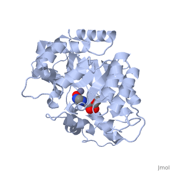
The crystal structure of a murine adenosine deaminase complexed with, 6-hydroxyl-1,6-dihydropurine ribonucleoside, a nearly ideal, transition-state analog, has been determined and refined at 2.4 angstrom, resolution. The structure is folded as an eight-stranded parallel, alpha/beta barrel with a deep pocket at the beta-barrel COOH-terminal end, wherein the inhibitor and a zinc are bound and completely sequestered. The, presence of the zinc cofactor and the precise structure of the bound, analog were not previously known. The 6R isomer of the analog is very, tightly held in place by the coordination of the 6-hydroxyl to the zinc, and the formation of nine hydrogen bonds. On the basis of the structure of, the complex a stereoselective addition-elimination or SN2 mechanism of the, enzyme is proposed with the zinc atom and the Glu and Asp residues playing, key roles. A molecular explanation of a hereditary disease caused by, several point mutations of an enzyme is also presented.
About this Structure
2ADA is a Single protein structure of sequence from Mus musculus with ZN and HPR as ligands. This structure superseeds the now removed PDB entry 1ADA. Active as Adenosine deaminase, with EC number 3.5.4.4 Full crystallographic information is available from OCA.
Reference
Atomic structure of adenosine deaminase complexed with a transition-state analog: understanding catalysis and immunodeficiency mutations., Wilson DK, Rudolph FB, Quiocho FA, Science. 1991 May 31;252(5010):1278-84. PMID:1925539
Page seeded by OCA on Wed Nov 21 08:03:45 2007
