This old version of Proteopedia is provided for student assignments while the new version is undergoing repairs. Content and edits done in this old version of Proteopedia after March 1, 2026 will eventually be lost when it is retired in about June of 2026.
Apply for new accounts at the new Proteopedia. Your logins will work in both the old and new versions.
2f4m
From Proteopedia
OCA (Talk | contribs)
(New page: 200px<br /><applet load="2f4m" size="450" color="white" frame="true" align="right" spinBox="true" caption="2f4m, resolution 1.85Å" /> '''The Mouse PNGase-HR2...)
Next diff →
Revision as of 08:14, 21 November 2007
|
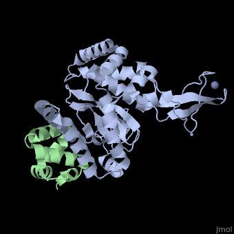
The Mouse PNGase-HR23 Complex Reveals a Complete Remodulation of the Protein-Protein Interface Compared to its Yeast Orthologs
Overview
Peptide N-glycanase removes N-linked oligosaccharides from misfolded, glycoproteins as part of the endoplasmic reticulum-associated degradation, pathway. This process involves the formation of a tight complex of peptide, N-glycanase with Rad23 in yeast and the orthologous HR23 proteins in, mammals. In addition to its function in endoplasmic reticulum-associated, degradation, HR23 is also involved in DNA repair, where it plays an, important role in damage recognition in complex with the xeroderma, pigmentosum group C protein. To characterize the dual role of HR23, we, have determined the high resolution crystal structure of the mouse peptide, N-glycanase catalytic core in complex with the xeroderma pigmentosum group, C binding domain from HR23B. Peptide N-glycanase features a large cleft, between its catalytic cysteine protease core and zinc binding domain., Opposite the zinc binding domain is the HR23B-interacting region, and, surprisingly, the complex interface is fundamentally different from the, orthologous yeast peptide N-glycanase-Rad23 complex. Different regions on, both proteins are involved in complex formation, revealing an amazing, degree of divergence in the interaction between two highly homologous, proteins. Furthermore, the mouse peptide N-glycanase-HR23B complex mimics, the interaction between xeroderma pigmentosum group C and HR23B, thereby, providing a first structural model of how the two proteins interact within, the nucleotide excision repair cascade in higher eukaryotes. The different, interaction interfaces of the xeroderma pigmentosum group C binding, domains in yeast and mammals suggest a co-evolution of the endoplasmic, reticulum-associated degradation and DNA repair pathways.
About this Structure
2F4M is a Protein complex structure of sequences from Mus musculus with ZN and CL as ligands. Active as Peptide-N(4)-(N-acetyl-beta-glucosaminyl)asparagine amidase, with EC number 3.5.1.52 Full crystallographic information is available from OCA.
Reference
Structure of the mouse peptide N-glycanase-HR23 complex suggests co-evolution of the endoplasmic reticulum-associated degradation and DNA repair pathways., Zhao G, Zhou X, Wang L, Li G, Kisker C, Lennarz WJ, Schindelin H, J Biol Chem. 2006 May 12;281(19):13751-61. Epub 2006 Feb 24. PMID:16500903
Page seeded by OCA on Wed Nov 21 10:21:47 2007
Categories: Mus musculus | Peptide-N(4)-(N-acetyl-beta-glucosaminyl)asparagine amidase | Protein complex | Kisker, C. | Lennarz, W.J. | Schindelin, H. | Wang, L. | Zhao, G. | Zhou, X. | CL | ZN | Glycoproteins | Nucleotide excision repair | Peptide:n-glycanase | Transglutaminase | Ubiquitin-dependent protein degradation
