X-ray crystallography
From Proteopedia
(Difference between revisions)
(→See Also - adding content) |
(→See Also - adding content) |
||
| Line 11: | Line 11: | ||
==See Also== | ==See Also== | ||
| + | *[http://www.pdb.org/pdb/static.do?p=general_information\about_pdb\nature_of_3d_structural_data.html Nature of 3D Structural Data] | ||
*[http://en.wikipedia.org/wiki/X-ray_crystallography X-ray Crystallography at Wikipedia] | *[http://en.wikipedia.org/wiki/X-ray_crystallography X-ray Crystallography at Wikipedia] | ||
Revision as of 21:01, 18 May 2009

|
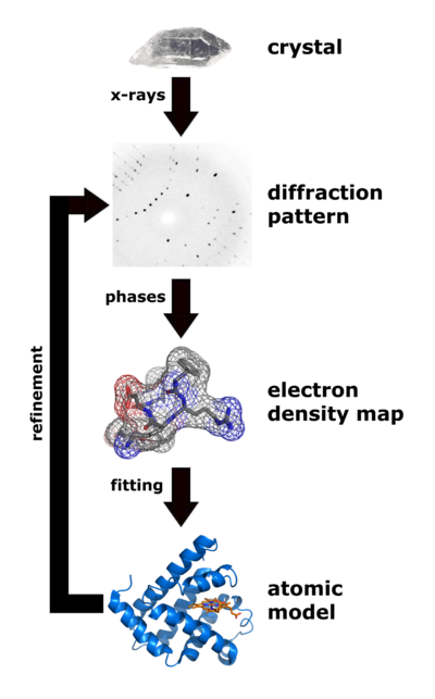
| Flow chart showing the major steps in X-ray protein crystallography. (Image from Wikimedia courtesy Thomas Splettstoesser. |
About 85% of the models (entries) in the World Wide Protein Data Bank were determined by X-ray crystallography. (Most of the remaining 15% were determined by solution nuclear magnetic resonance.) Protein crystallography remains very difficult, despite many recent advances. For every new protein sequence targeted for X-ray crystallography, about one in twenty is solved[1][2]. Publication of solved structures involves depositing an atomic coordinate file (PDB file) in the World Wide Protein Data Bank.
