We apologize for Proteopedia being slow to respond. For the past two years, a new implementation of Proteopedia has been being built. Soon, it will replace this 18-year old system. All existing content will be moved to the new system at a date that will be announced here.
Tedsandbox
From Proteopedia
(Difference between revisions)
| Line 47: | Line 47: | ||
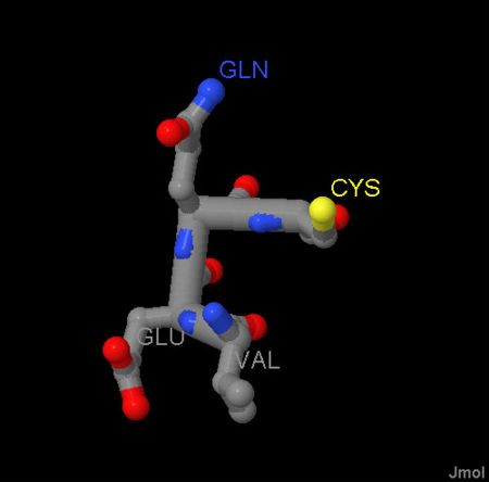
rect 100 419 167 305 [http://en.wikipedia.org/wiki/Glutamic_acid] | rect 100 419 167 305 [http://en.wikipedia.org/wiki/Glutamic_acid] | ||
circle 235 373 60 [http://en.wikipedia.org/wiki/Valine] | circle 235 373 60 [http://en.wikipedia.org/wiki/Valine] | ||
| - | rect 250 243 350 200[http://en.wikipedia.org/wiki/cystine] | + | rect 250 243 350 200 [http://en.wikipedia.org/wiki/cystine] |
| - | circle 188 158 55 [ | + | circle 188 158 55 [http://en.wikipedia.org/wiki/Glycine] |
# A comment, this line is ignored. | # A comment, this line is ignored. | ||
# desc bottom-left | # desc bottom-left | ||
</imagemap> | </imagemap> | ||
Revision as of 10:20, 15 July 2009
I think a line at the top looks nice
|
Need more help?
Need more help?
Perhaps it was confusing because you are not used to seeing this protein as a dimer.
Last look.
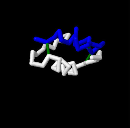
Click on the disulfide bonds or the white alpha helix for more information.
Click on the amino acids for more information.


