File list
From Proteopedia
| Name | User | Size | Description | |
|---|---|---|---|---|
| 08:06, 10 February 2009 | Coenzyme_Q10.pdb (file) | Ralf Stephan | 14 KB | (CORINA model of coenzyme Q10) |
| 09:49, 9 February 2009 | Dicoumarol.pdb (file) | Ralf Stephan | 3 KB | (CORINA solution of dicoumarol structure) |
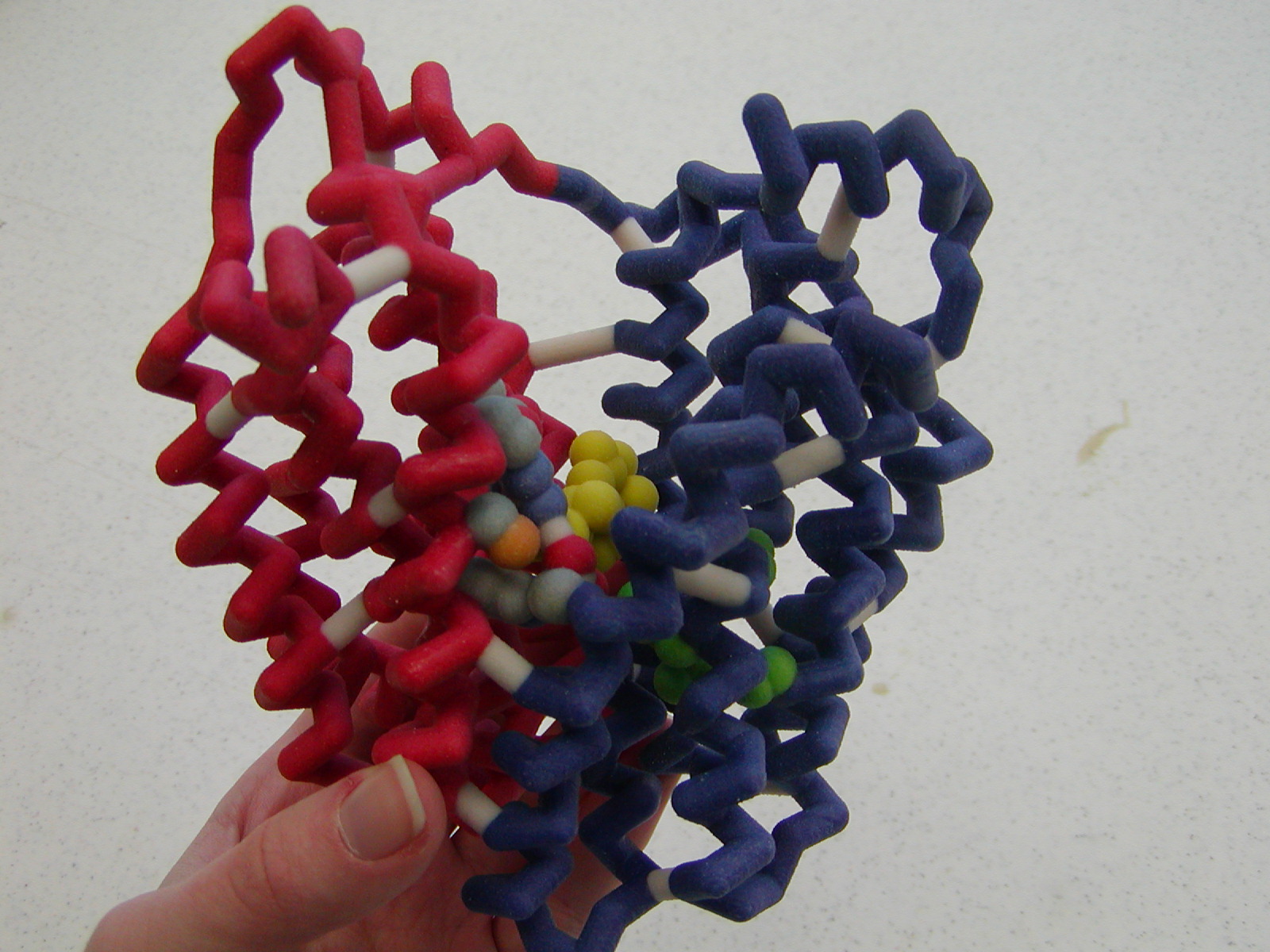
| 10:53, 8 February 2009 | ACHE4.pdb (file) | Alexander Berchansky | 1.36 MB | (2ace + 1gqr + 1gqs + 1vxr) |
| 10:33, 8 February 2009 | Acetylcholine.pdb (file) | Ralf Stephan | 2 KB | (CORINA solution of acetylcholine) |
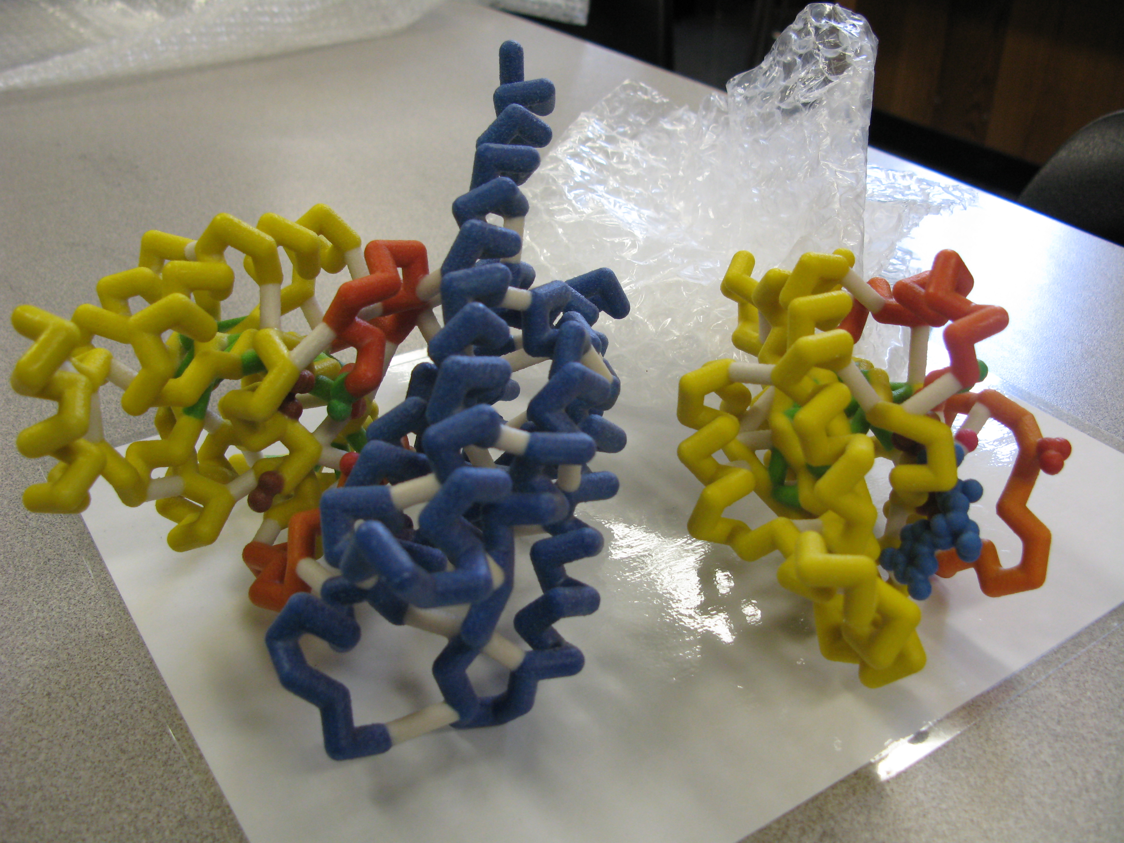
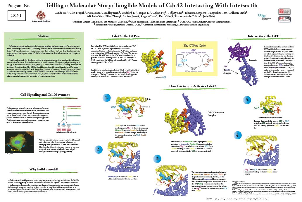
| 09:27, 8 February 2009 | Actual_3d_models.JPG (file) | Elizaveta Sergeeva | 3.48 MB | (Actual 3D models of complexes cdc42 with GDP and Intersectin (DH domain) with cdc42) |
| 09:16, 8 February 2009 | Lincoln_Team_2008.JPG (file) | Elizaveta Sergeeva | 4.08 MB | (Lincoln Team 2008) |
| 09:01, 8 February 2009 | Poster_picture.jpg (file) | Elizaveta Sergeeva | 143 KB | (Lincoln High School poster of the project.) |
| 11:11, 7 February 2009 | Filopodia.jpg (file) | Elizaveta Sergeeva | 40 KB | (Lim Lab, UCSF) |
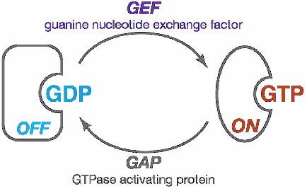
| 11:02, 7 February 2009 | GDP_to_GTP.jpg (file) | Elizaveta Sergeeva | 17 KB | (How GDP changes to GTP) |
| 08:49, 7 February 2009 | 1an0.pdb (file) | Elizaveta Sergeeva | 283 KB | (Cdc42 with GDP) |
| 08:26, 7 February 2009 | INTERSECTIN.MOL (file) | Elizaveta Sergeeva | 30 KB | (Complex Intersectin and Cdc42) |
| 00:42, 7 February 2009 | Heckert_GNU_white.png (file) | Angel Herraez | 40 KB | (The GNU logo. Source: WikiMedia Commons http://commons.wikimedia.org/wiki/File:Heckert_GNU_white.png Copyleft: This work of art is free; you can redistribute it and/or modify it according to terms of the Free Art License. You will find a specimen of this) |
| 00:12, 7 February 2009 | Cc-pd-40x40.gif (file) | Angel Herraez | 582 B | (Creative Commons logo for Public Domain. Valid use of the logo is specified in Creative Commons Trademark policy, http://creativecommons.org/policies ) |
| 23:56, 6 February 2009 | Cc-by-sa-88x31.png (file) | Angel Herraez | 5 KB | (Creative Commons logo for Attribution-Share Alike License. Valid use of the logo is specified in Creative Commons Trademark policy, http://creativecommons.org/policies ) |
| 23:40, 6 February 2009 | Cc-by-88x31.png (file) | Angel Herraez | 5 KB | (Creative Commons logo for Attribution License. Valid use of the logo is specified in Creative Commons Trademark policy, http://creativecommons.org/policies ) |
| 15:17, 6 February 2009 | 1ea5_biol_unit.pdb (file) | Eran Hodis | 672 KB | (Biological unit of 1ea5 as predicted by PISA ) |
| 16:17, 4 February 2009 | Zf_assort.jpg (file) | Savannah Anderson | 56 KB | (This is an assortment of different representations of zinc finger models. Some of the visible variations include wireframe, spacefill, and backbone representations.) |
| 11:10, 3 February 2009 | 7_scrolling.swf (file) | Jaime Prilusky | 25 KB | (retrieved as a sample from http://www.adobe.com/devnet/flash/samples/interactivity_7/index.html) |
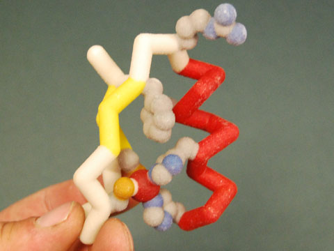
| 21:34, 2 February 2009 | Zf_toober.jpg (file) | Savannah Anderson | 34 KB | (This is a model of a zinc finger created using 'mini-toobers.' The Residues are shown in CPK color format and the zinc atom is shown in green.) |
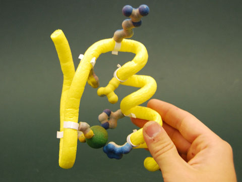
| 21:24, 2 February 2009 | Zf_rpmodel.jpg (file) | Savannah Anderson | 40 KB | (This is a physical model of a zinc finger generated from the PDB file 1ZAA. The model was designed and built by the MSOE Center for BioMolecular modeling using rapid prototyping technology. In this model, the alpha helix is depicted in red, while the be) |
| 05:55, 1 February 2009 | 2az0WithPolyview3D.png (file) | Wayne Decatur | 309 KB | (2az0 with Polyview-3D Settings used: Data source: PDB code=2az0 Initial rotation around X axis: -85.5 Initial rotation around Y axis: -77.2 Initial rotation around Z axis: 38.1 Rendering program: PyMol Type of image: Static slide Background color Red fac) |
| 14:58, 31 January 2009 | Alg44_PSEAE_MEXA_1vf7.pdb (file) | Tilman Schirmer | 125 KB | (Alg44, Mexa domain, modeled on 1vf7 (HHPred)) |
| 11:23, 31 January 2009 | Alg44_PSEAE_PILZ_2gjg_super2rde.pdb (file) | Tilman Schirmer | 64 KB | (Homology model of the PilZ domain of Alg44_PSEAE modeled (HHpred) with 2gjgas template moved to superimpose 2rde) |
| 21:06, 30 January 2009 | 1kyo_cn.jvxl (file) | Karl Oberholser | 18 KB | (Data file generated with Jmol commands select :c, :n; isosurface surface_cn minset 1000 pocket cavity 1.2 using 1kyo.pdb. ) |
| 15:03, 30 January 2009 | CC0857_GGDEF_1w25.pdb (file) | Tilman Schirmer | 107 KB | (Homology model of GGDEF domain of CC0857, Template 1w25) |
| 14:35, 30 January 2009 | CC0740_GGDEF_1w25.pdb (file) | Tilman Schirmer | 109 KB | (Homology model of GGDEF domain of CC0740, Template 1w25) |
| 13:23, 30 January 2009 | CC0655_GGDEF_1w25.pdb (file) | Tilman Schirmer | 110 KB | (Homology model of GGDEF domain of CC0655, Template 1w25) |
| 12:45, 30 January 2009 | CC0091_GGDEF_1w25.pdb (file) | Tilman Schirmer | 108 KB | (Homology model of GGDEF domain of CC0091, Template 1w25) |
| 11:30, 30 January 2009 | PdeA_CAUCR_GGDEF_1w25.pdb (file) | Tilman Schirmer | 92 KB | (Homology model of GGDEF domain of PdeA_CAUCR, Template 1w25) |
| 10:36, 30 January 2009 | DgcB_CAUCR_1w25.pdb (file) | Tilman Schirmer | 116 KB | (Homology model of GGDEF domain of DgcB_CAUCR, Template 1w25) |
| 22:48, 29 January 2009 | 1jgo1giy.gz.pdb (file) | Wayne Decatur | 296 KB | (pdb files 1jgo and 1giy as one gzipped file for use with jmol See Yusupov MM, Yusupova GZ, Baucom A, Lieberman K, Earnest TN, Cate JH, and Noller HF. 2001. Crystal structure of the ribosome at 5.5 Å resolution. Science 292:883-896. Epub 2001 Mar 29. an) |
| 14:22, 29 January 2009 | PopA_CAUCR_GGDEF_1w25.pdb (file) | Tilman Schirmer | 96 KB | (Homology model of GGDEF domain of PopA_CAUCR, Template 1w25) |
| 12:28, 29 January 2009 | DgcA_CAUCR_GGDEF_1w25.pdb (file) | Tilman Schirmer | 105 KB | (Homology model of GGDEF domain of DgcA_CAUCR, template 1w25) |
| 08:14, 28 January 2009 | PopA_1w25.pdb (file) | Tilman Schirmer | 219 KB | (Homology model of PopA_CAUCR based on template 1w25 (PleD_CAUCR). Ludwig Zumthor (2008) Master Thesis, University of Basel, Switzerland) |
| 06:40, 28 January 2009 | 2zwh.png (file) | OCA | 215 KB | (Source Jena Library http://www.fli-leibniz.de/IMAGE.html) |
| 09:47, 27 January 2009 | 1w25_small.png (file) | Tilman Schirmer | 67 KB | |
| 08:44, 21 January 2009 | 3d06.jpg (file) | OCA | 36 KB | (Source PDB http://www.rscb.org/pdb) |
| 21:08, 15 January 2009 | Smart_Teams_photo_5.jpg (file) | Mark Hoelzer | 43 KB | (SMART Teams run by the Center for BioMolecular Modeling. ) |
| 21:00, 15 January 2009 | Smart_Team_photo_4.jpg (file) | Mark Hoelzer | 22 KB | (SMART Teams run by the Center for BioMolecular Modeling. ) |
| 21:00, 15 January 2009 | Smart_Team_photo_3.jpg (file) | Mark Hoelzer | 16 KB | (SMART Teams run by the Center for BioMolecular Modeling. ) |
| 20:59, 15 January 2009 | Smart_team_2.jpg (file) | Mark Hoelzer | 23 KB | (SMART Teams run by the Center for BioMolecular Modeling. ) |
| 20:39, 15 January 2009 | SMART_Teams_photo_1.jpg (file) | Mark Hoelzer | 33 KB | (SMART Teams run by the Center for BioMolecular Modeling.) |
| 16:33, 15 January 2009 | Lacpermmodel.jpg (file) | Savannah Anderson | 415 KB | (This is an image of a physical model of Lactose Permease created by the Center for BioMolecular Modeling at the Milwaukee School of Engineering. The model was based off the PDB file 1pv7.) |
| 13:26, 11 January 2009 | ACHE3.pdb (file) | Alexander Berchansky | 1.03 MB | (1e3q + 1acl + 1eve) |
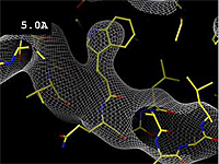
| 22:57, 2 January 2009 | Resolution-holton-5.0.png (file) | Eric Martz | 55 KB | (Still from Image:Resolution holton.mpeg) |
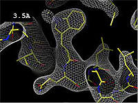
| 22:57, 2 January 2009 | Resolution-holton-3.5.png (file) | Eric Martz | 59 KB | (Still from Image:Resolution holton.mpeg) |
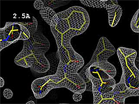
| 22:57, 2 January 2009 | Resolution-holton-2.5.png (file) | Eric Martz | 62 KB | (Still from Image:Resolution holton.mpeg) |
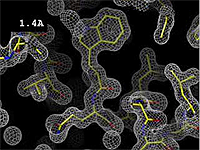
| 22:56, 2 January 2009 | Resolution-holton-1.4.png (file) | Eric Martz | 64 KB | (Still from Image:Resolution holton.mpeg) |
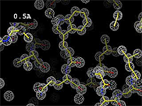
| 22:56, 2 January 2009 | Resolution-holton-0.5.png (file) | Eric Martz | 54 KB | (Still from Image:Resolution holton.mpeg) |
| 16:09, 2 January 2009 | Resolution_holton.mpeg (file) | Eric Martz | 2.12 MB | (Movie showing relation between resolution, the electron density map, and the atomic model, prepared by James Holton at the Advanced Light Source of the Berkeley Laboratory at the University of California. Holton gave explicit permission to use this movie ) |
First page |
Previous page |
Next page |
Last page |
