File list
From Proteopedia
| Name | User | Size | Description | |
|---|---|---|---|---|
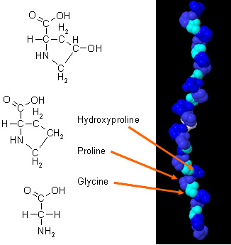
| 01:46, 27 March 2010 | Collagen_(alpha_chain).jpg (file) | Daman K. Kandola | 31 KB | (This picture illustrates the glycine, proline and hydroxyproline residues present in collagen.) |
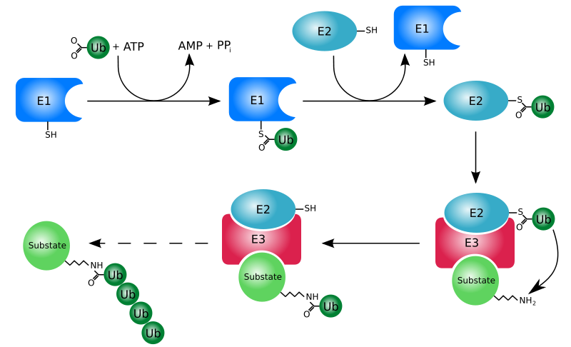
| 21:25, 26 March 2010 | Ubq_pathway.png (file) | Jaclyn Gordon | 77 KB | |
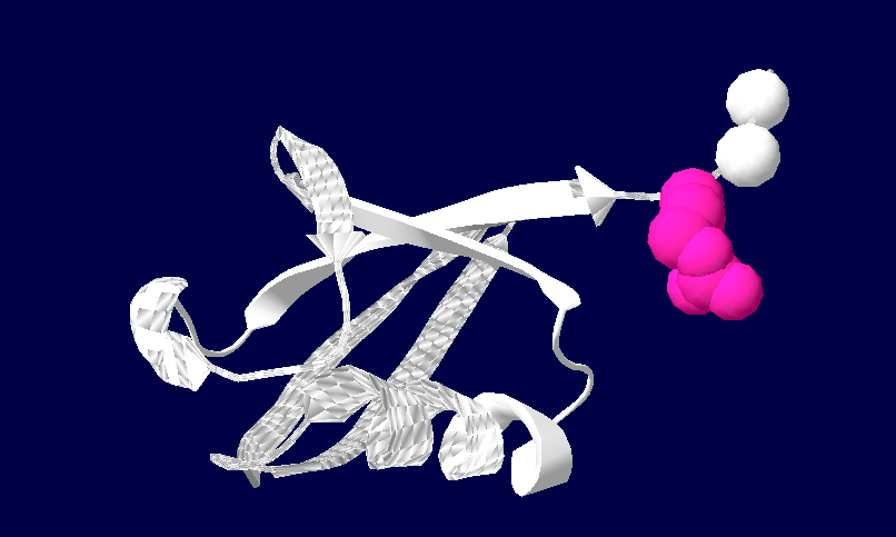
| 21:09, 26 March 2010 | 1ubiq.png (file) | Jaclyn Gordon | 69 KB | |
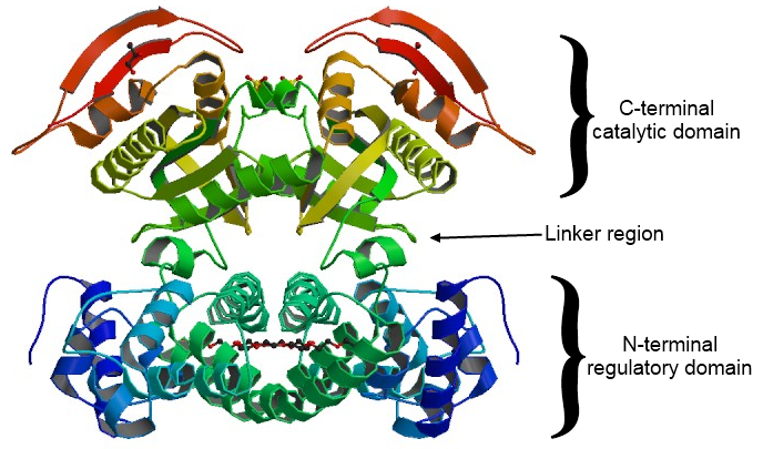
| 20:11, 26 March 2010 | Dimer2.png (file) | Travis Eyford | 322 KB | |
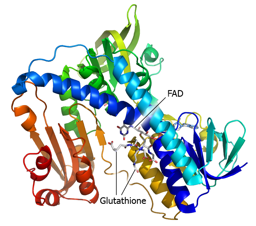
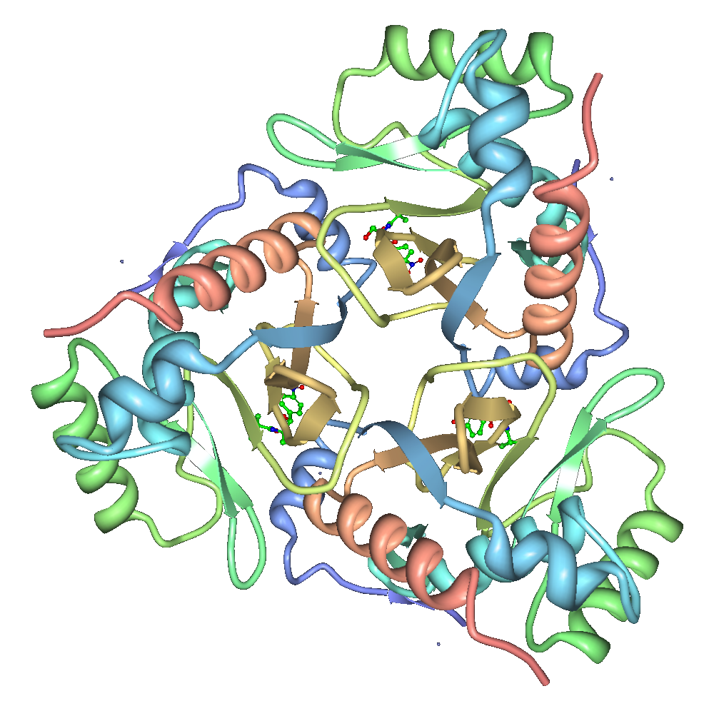
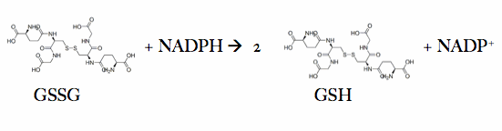
| 19:58, 26 March 2010 | Glutathione_reductase.png (file) | Josina Rhebergen | 305 KB | |
| 19:32, 26 March 2010 | 2HU4monomer.png (file) | Andrea Gorrell | 131 KB | |
| 19:21, 26 March 2010 | Catalytic_domain_of_YopH.gif (file) | Cara Halseth | 14 KB | (The residue of the catalytic domain of YopH closest to the N-terminal is coloured in blue. The C-terminal residue is red, and the phosphate-binding loop in the active site is green. Phosphopeptide ligands are shown in purple ) |
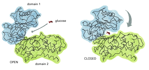
| 13:09, 26 March 2010 | Glucosebinding.jpg (file) | Amanda Lam | 75 KB | |
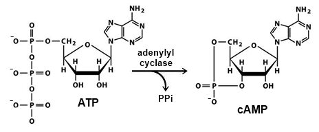
| 09:04, 26 March 2010 | CAMP_synthesis.png (file) | Travis Eyford | 23 KB | (cAMP and PPi formation from ATP cayalyzed by adenylyl cyclase) |
| 07:46, 26 March 2010 | 512px-Acetyl-CoA-2D.svg.png (file) | Anthony Daniele | 8 KB | (Structure of acetyl-CoA) |
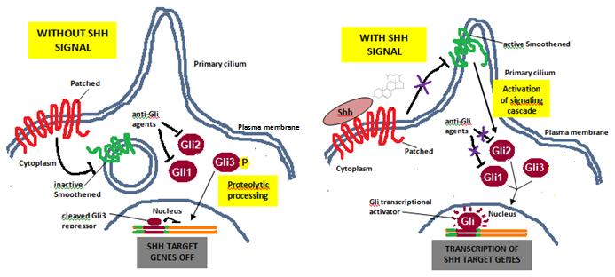
| 07:42, 26 March 2010 | SHH_SIGNALING_PATHWAY.jpg (file) | Randi Woodbeck | 39 KB | (Sonic hedgehog signaling pathway. ) |
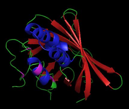
| 07:22, 26 March 2010 | 1JUN.png (file) | Andrew Rebeyka | 45 KB | (cfvvv) |
| 03:57, 26 March 2010 | 1yr2_ribbon2.png (file) | Stacey Shantz | 77 KB | (Image of 1yr2 in ribbon format with linkages between domains highlighted.) |
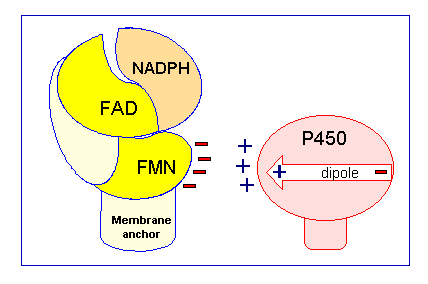
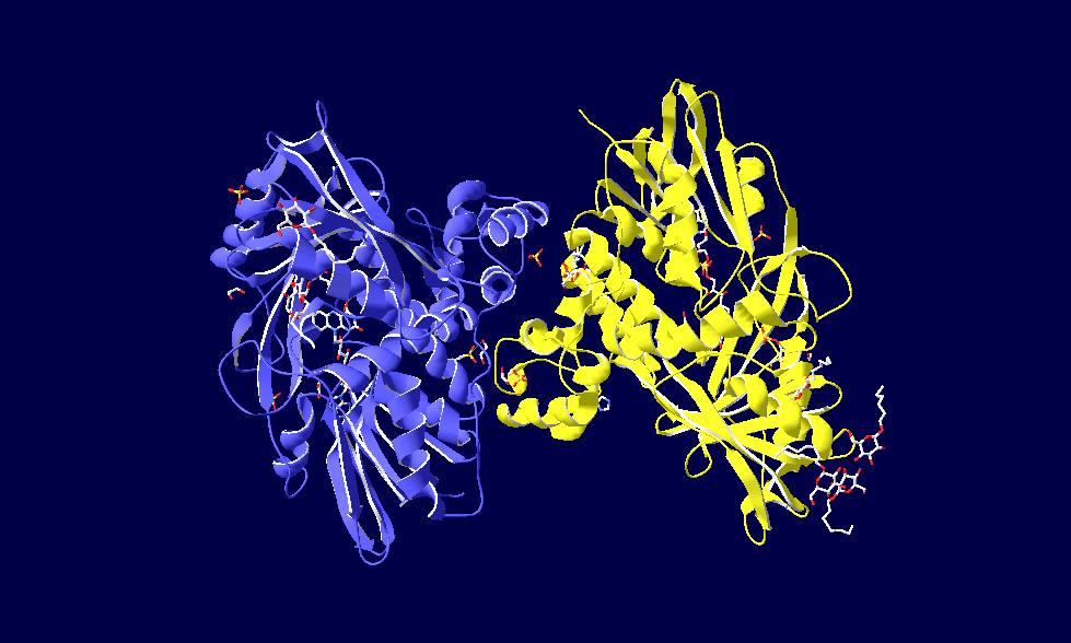
| 00:19, 26 March 2010 | CPR_chargepair.gif (file) | Taya O'Neill | 6 KB | (This is a picture of CYPOR interacting with Cyt P450 in cartoon form) |
| 22:02, 25 March 2010 | FINAL.png (file) | Andrea Gorrell | 146 KB | |
| 21:57, 25 March 2010 | Sidechain_import.JPG (file) | Christine Brown | 8 KB | (this is an image of teh side chains involved in binding) |
| 21:55, 25 March 2010 | Ribbon_import.JPG (file) | Christine Brown | 7 KB | (this is an imapge in ribbon form of important residues involved in binding) |
| 21:32, 25 March 2010 | SOD1.png (file) | Jordan Schibli | 58 KB | (Cu-Zn Superoxide dismutase chain) |
| 13:02, 25 March 2010 | Crdes2.pdb (file) | Alexander Berchansky | 908 KB | (Alignment 2rkx, 3iio, 3iiv, 3iip) |
| 00:32, 25 March 2010 | N_Teeerminal_Hydrophobic.png (file) | Elyse Yaremco | 35 KB | |
| 22:26, 24 March 2010 | Full_structure.pdb (file) | Travis Eyford | 246 KB | (full structure- 1ab8) |
| 16:24, 24 March 2010 | Structure_trihelix_barrel.jpg (file) | James Jones | 17 KB | (Cropped PyMol image of 1vpr highlighting the tri-helix and beta-barrel structure. Note that residues implied in pH-depentant activity regulation are coloured pink. ) |
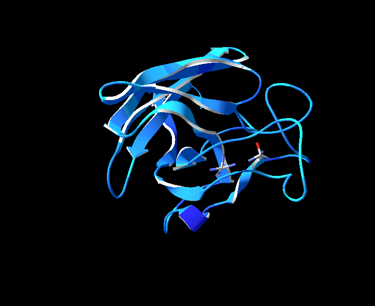
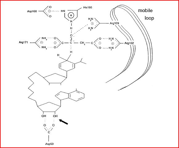
| 03:24, 24 March 2010 | 1vc2_image.png (file) | Satvir Gill | 204 KB | (This is an illustration of certain residues in the active site of 1vc2.) |
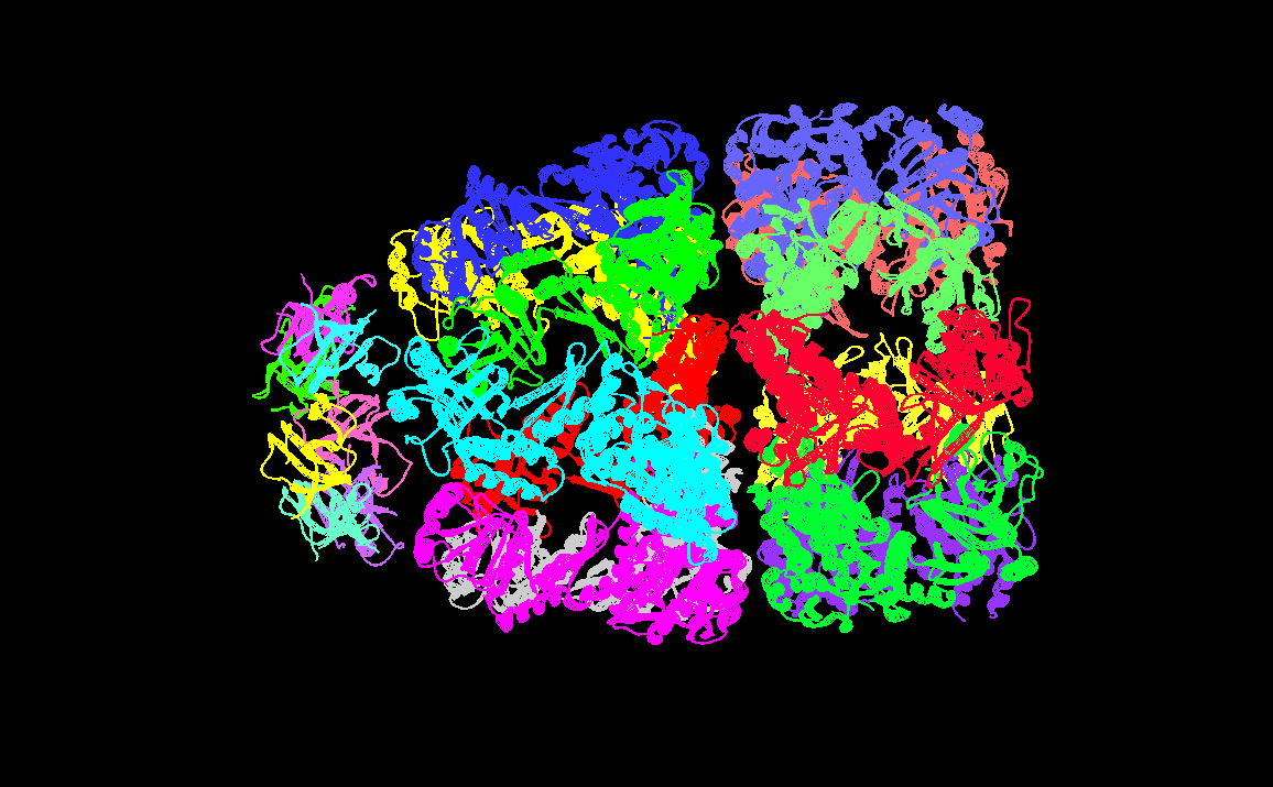
| 22:26, 23 March 2010 | GroELGroES.png (file) | Andrea Gorrell | 61 KB | |
| 21:26, 23 March 2010 | Fig2.gif (file) | Jordan Schibli | 1 KB | (Superoxide dismutase reaction) |
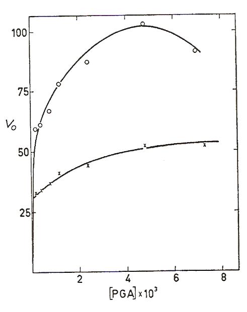
| 21:11, 23 March 2010 | Enolase_kinetics.jpeg (file) | Cory Tiedeman | 19 KB | (Shows the V vs. [S] graph of enolase with two different concentrations of Mg2+. The substrate, PGA, is actually 2-PG. The upper curve has an Mg2+ concentration of 10^-3 M and the lower curve has an Mg2+ concentration of 10^-2 M.) |
| 20:45, 23 March 2010 | Superoxide_dismutase.png (file) | Jordan Schibli | 92 KB | |
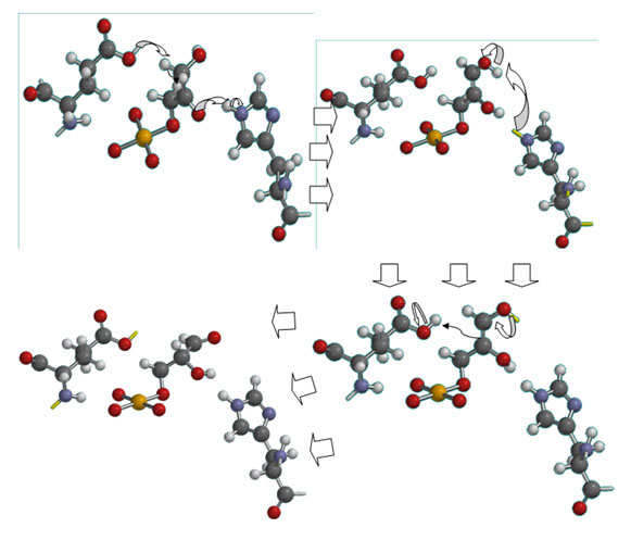
| 12:56, 23 March 2010 | Ckrenkmechanism.jpg (file) | Christian Krenk | 71 KB | (Triose phosphate isomerase mechanism) |
| 02:45, 23 March 2010 | 1CZA.pdb.gz (file) | Amanda Lam | 166 KB | (This is my protein) |
| 02:38, 23 March 2010 | 1Z82.pdb.gz (file) | Indu Toora | 116 KB | |
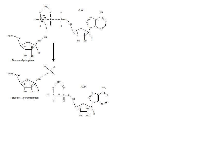
| 14:11, 22 March 2010 | PFK_mech.JPG (file) | Zach Westrick | 23 KB | |
| 07:26, 22 March 2010 | Malate_Dehydrogenase_Active_Site.JPG (file) | Jake Ezell | 37 KB | (A picture of the mechanism of malate dehydrogenase.) |
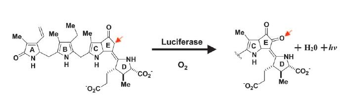
| 05:51, 22 March 2010 | Luciferase_reaction.jpg (file) | James Jones | 14 KB | (The reaction of dinoflagellate luciferase to produce light. Image courtesy of L. Wayne Schultz.) |
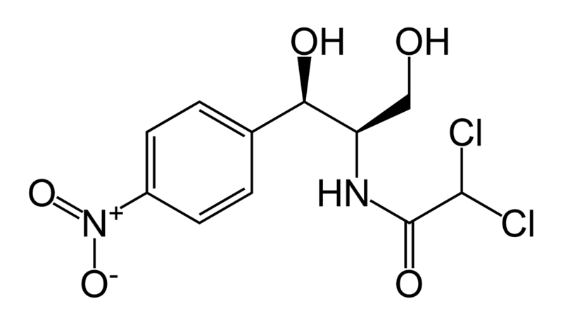
| 18:45, 21 March 2010 | Chloramphenicol.png (file) | Anthony Daniele | 40 KB | (Chloramphenicol chemical structure) |
| 18:32, 21 March 2010 | Chloramphenicol_acetyltransferase_3CLA_transparent.png (file) | Anthony Daniele | 513 KB | (Cartoon representation of the enzyme CAT III.) |
| 20:56, 20 March 2010 | Eric_Fadel_OIST_cropped.jpg (file) | Eric Martz | 105 KB | |
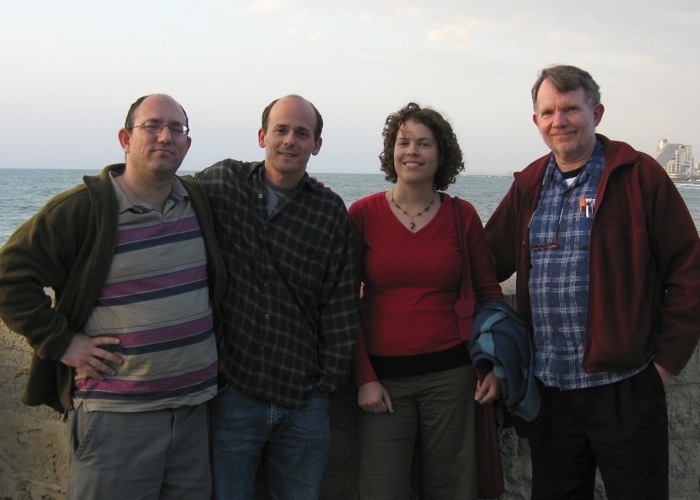
| 20:14, 20 March 2010 | Fabian_Tal_Elana_Eric_cropped_2008.jpg (file) | Eric Martz | 80 KB | |
| 20:09, 20 March 2010 | Nir_Ben-Tal_and_Eric_cropped_2008.jpg (file) | Eric Martz | 45 KB | |
| 19:51, 20 March 2010 | Jaim_Eran_Eric_2008_small_adj.jpg (file) | Eric Martz | 150 KB | |
| 19:26, 20 March 2010 | Joel_Jaim_Eran_Eric_Pub_2008_small_adj.jpg (file) | Eric Martz | 179 KB | |
| 18:54, 20 March 2010 | Tom_and_eric_and_slide_UMBC_2009.jpg (file) | Eric Martz | 74 KB | |
| 18:29, 20 March 2010 | Namba_Team_ProteopediaShirts.jpg (file) | Eric Martz | 111 KB | |
| 05:48, 19 March 2010 | Glutathione_reductase_mechanism.gif (file) | Josina Rhebergen | 7 KB | |
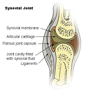
| 01:23, 19 March 2010 | Illu_synovial_joint.jpg (file) | Shelly Huebert | 33 KB | |
| 12:54, 18 March 2010 | Crdes1.pdb (file) | Alexander Berchansky | 264 KB | (2rkx+) |
| 00:22, 18 March 2010 | Ligand1.PNG (file) | Shannon King | 24 KB | |
| 17:44, 17 March 2010 | 1K4C_tetramer.pdb (file) | Tilman Schirmer | 1.42 MB | (Biological unit (tetramer) of 1K4C (obtained from the PDB).) |
| 06:53, 17 March 2010 | 3l2q.png (file) | OCA | 219 KB | (Source Jena Library http://www.fli-leibniz.de/IMAGE.html) |
| 15:17, 16 March 2010 | Trans-cis-peptide-bond.png (file) | Tilman Schirmer | 28 KB | (Schematics of trans and of cis peptide bond) |
| 12:47, 16 March 2010 | MP_relenza03.spt (file) | Eric Martz | 2 KB |
First page |
Previous page |
Next page |
Last page |
