Monoclonal Antibody
From Proteopedia

Monoclonal antibodies are immunoglobulins produced by a single clone of cells and therefore a single pure homogeneous type of antibody. For structural information on immunoglobulins, see: Antibody The development of monoclonal antibodies has revolutionized much of the science world both in the lab and in the medical world in treating cancer and autoimmune disorders.
Brief History
Nobel Laureate Paul Ehrlich proposed the idea of a “magic bullet” at the beginning of the 20th century by postulating that if a compound could be made that selectively targets a disease causing organism, then toxins could be delivered to selectively kill any cell. He proposed that this could be a cure for nearly any disease. The monoclonal antibody is this “magic bullet,” and while they are extremely selective and effective, monoclonal antibodies have limitations. Georges Kholer, Cesar Milstein, and Niels kaj Jerne developed the monoclonal antibody and shared the Nobel Prize in Medicine in 1984 for the discovery.
Original Monoclonal Antibody Technology
When an organism is exposed to an antigen, the immune system stages a complex immunological response. One such response is the activation of B-cells and subsequent release of antibodies. To any single antigen, thousands of different B-cells can be activated by their binding different epitopes on that antigen. When these B-cells subsequently mature into antibody releasing plasma cells, thousands of different antibodies are released into the blood binding and removing the invading antigen. The body creates these “polyclonal” antibodies to guarantee antigens are bound multiple times by antibodies to expedite their removal and serves as a redundant form of immunological security in case some of the antibodies produced are faulty. [1] In order to be a useful tool in research and medicine however, a single, monoclonal antibody must be isolated.
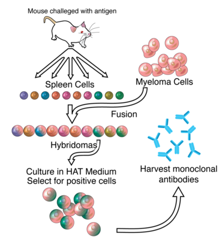
Monoclonal antibody production has changed drastically since Kholer and Milstein first did it in 1975. A brief description of their process however is insightful. First an antigen of choice is injected into a mouse and after 10 days, a sample of B-cells is extracted from the spleen of the mouse. These cells are added to a culture of myeloma cells, which are immortal cancer cells, to form hybridomas, cells formed by the fusion of a B-cell and myeloma cells. [2] Next, the hybridomas undergo selection hypozanthine-aminopterin-thimine (HAT) medium, in which only those cells that have successfully formed fusions will survive indefinitely. The hybridomas are then cultured and screened after doing SDS-PAGE and Western blots to identify those hybridomas creating the desired antibody. These hybridomas are immortal and can produce “murine” antibodies nearly indefinitely. [3]
Newer Monoclonal Technology
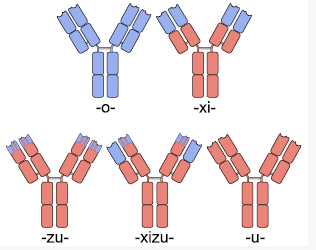
Early forms of monoclonal antibodies (murine antibodies) were problematic for therapeutic use because they were mouse antibodies, not human antibodies. When injected into humans, the antibodies would either be rapidly cleared from the body or worse, result in systemic inflammatory effects that were harmful. The human immune system recognized these mouse antibodies as foreign pathogens and rapidly produced human anti-mouse antibodies and unleashed the immune system on these invading pathogens. [4] To avoid this immune response, antibodies that the body recognized as native had to be created. Murine antibodies typically have a “mo” before the “mab” in their name, as is the case with Tositumomab (Marketed as Bexxar by GlaxoSmithKline), a drug used to treat lymphoma.
|
Chimeric Antibodies
One solution to this problem was the creation of chimeric antibodies. Chimeric anibodies are antibodies that have the , but have . These are created by fusing the Fab region coding DNA extracted from mice B-cells as above to human antibody DNA that only codes for the Fc region. Since it is the Fc region of an antibody that is glycosylated and used by the human immune system to determine self vs non-self, and the Fc region in chimeric antibodies is human, these chimeric antibodies typically cause a minimal immune response against them and thus have a longer half life. [5]. Chimeric antibodies typically have a “xi” before the “mab” at the end of their name to indicate their chain type, as is the case with “abciximab” (ReoPro), “cutximab” (Erbitux), or (Rituxan).
These chimeric antibodies can be further humanized through mutagenesis to the Fab sequence (excluding the ), identifying those parts which differ in humans and mutating them. This is a difficult process, but can create antibodies that are unrecognizable from human antibodies at the amino acid level. [6]. To avoid the added mutagenesis step, insertion of only the CDRs from mice into a full human antibody scaffold has been used to create humanized antibodies.[7] Humanized antibodies typically have a “zu” before the “mab” to indicate their chain type. The drug Alemtuzumab (marketed by Genzyme for the treatment of leukemia) is an example of a humanized antibody with foreign CDR regions.
Human Antibody Production & Phage Display
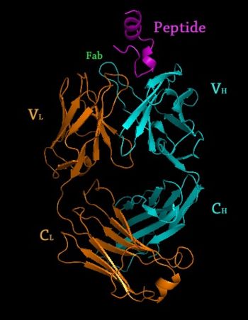
The major drawback with chimeric antibody production is it still involves using rodents as the original source of antigen binding, and requires significant effort to identify, purify and create the fusion antibody. Applying a technique called phage display to monoclonal antibody production expedites this entire process. In phage display, DNA coding the binding region of the antibody is ligated into the genes of a phage which code for proteins that are expressed on the protein coat of the virus. This phage gene hybrid is then transformed into bacterial cells. The virus hijacks the bacterial replication machinery, producing vast quantities of phage. By using many DNA fragments that are slightly different, a library of slightly different antibodies can be expressed on the outside of these phages, one type of antibody per phage. These antibody covered phages can then be selected for by running them through a column with the protein target immobilized on the column. Once the phages that bind the target protein well are identified, they can be used to reinfect bacterial cells and to isolate the DNA of antibodies that code for those antibodies with the greatest affinity for the target. Once the best antibodies are identified, they can be combined with human antibody scaffolds or human Fc regions as above to create humanized antibodies. [8] [9]Antibodies created through phage display have a “u” before the “mab” in their name to indicate their chain origins as is the case for Adalimumab (HUMIRA) Template:STRUCTURE 1l6x
Glycosylation of Monoclonal Antibodies
Once an antibody gene is designed to contain the ideal sequence, the antibody then must be mass produced. An ideal system of production would be in bacterial cells because of the rapidity with which they replicate and ease of use. Unfortunately, bacterial cells do not glycosylate proteins the same way mammalian cells do. Since is critical for the immune system to recognize a protein as native, thus allowing the protein to stay in circulation in the human body without creating an immune response, antibody production had to be completed in cells that glycosylate the antibody appropriately. Chinese Hamster Ovary (CHO) cells are one type of cell that is able to glycosylate antibodies appropriately, but they are difficult to work with and relatively expensive at an industrial scale. Regardless, they are the most commonly used mammalian hosts for industrial production of recombinant protein therapeutics. [10] More recently, work by Dr. Tillman Gerngross, et al. have demonstrated that yeast can be genetically manipulated to produce human glycosylation on expressed antibodies. This is an important discovery because yeast replicate much faster than CHO cells, making them more ideal as hosts for industrial scale production of therapeutic proteins. [11] Gerngross’s startup company GlycoFi, which was based around this platform was purchased by Merck in 2006 and the technology is being employed in Merck’s pipeline. Gerngross’s new company, [www.adimab.com Adimab], also uses similar technology to produce human glycosylated proteins for the pharmaceutical industry.
Monoclonal Antibody Uses
Laboratory Research
Monoclonal antibodies have become a critical tool in most biochemistry labs. They are essential components in immunofluorescence experiments as well as in Western Blot and immuno dot blot tests to detect protein on a membrane.
Medical Applications
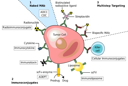
Monoclonal antibodies have been utilized to create four major classes of drugs. The first activate the bodies own immune system. An example of this is Rituximab (Marketed as Rituxan by Roche and Genentech). to CD20 surface proteins on B-Cells and illicits natural immune responses to these cancerous cells via ADCC, activating cellular apoptosis, or by attracting compliment proteins which kill the cell via the compliment pathway. [12] Another example of this type of monoclonal antibody drug is Infliximab (Marketed as [www.remicade.com/ Remicade] by Merck and the best selling Monoclonal therapeutic in 2008 with over $6 billion in sales). This class of antibodies also includes those antibodies that simply bind a target, disrupting a key mechanism in the diseased cells.
The second class of monoclonal antibody drugs are those conjugated to a disruptive compound. One example of this is used in Radioimmunotherapy (RIT). By conjugating a radioactive isotope to a murine antibody, targeted immunotherapy is possible. The antibody binds to the targeted cancer cell and the cell is damaged or destroyed by emitted beta particles. Murine antibodies are used because they are cleared from the body rapidly so as to avoid extensive radiation damage from the drug. This is the mechanism used by Tositumomab (Bexxar) which uses radioactive Iodine-131. [13] Also within this class of antibodies are those antibodies conjugated to a chemotherapy drug. An example of this type would be Gemtuzumab (marketed as Mylotarg by Wyeth until June 2010, when it was removed from the market due to inefficacy and safety concerns)
The Third class of monoclonal antibody drugs are those conjugated to a drug activating enzyme also known as Antibody-Directed Enzyme Prodrug Therapy (ADEPT). In this system, an enzyme that is able to activate a drug is attached to an antibody with specificity toward a target of choice. After injection of the antibody/enzyme compound and ample time for the antibodies to bind the target, a prodrug that is inert until activated by the enzyme is injected. This prodrug stays inert until it comes into contact with the antibody/enzyme compound bound to the target, whereupon the prodrug is activated and is able to attack cells in the local environment, which presumably would be populated by the infected target cells. No ADEPT system drugs have made it through the clinic yet, but the class holds promise. [14] [15]
The Final class of monoclonal antibody medical uses are those conjugated to liposomes or another form of nanotechnology drug delivery system. By attaching antibodies to the outside of a nanosized drug delivery device, large quantities of therapeutic drug can be delivered to a targeted environment. Many new nanotech devices including liposomes, nanotubes and other such containers have been developed and have demonstrated improved specificity for delivery existing drug compounds. [16]
Brand New Research
Here are some new tidbits of interesting information regarding monoclonal Antibodies
Lippow et al. have developed a computational method for improving antibody binding to targets. They were able to increase cetuximab (Erbitux) affinity 10 fold and comparable improvements in bevacizumab (Avastin), which disrupts Vascular Endothelial Growth Factor from binding VEGFR. [17]
Gribenko et al. developed a computation method for making any antibody more stable in the body by making specific changes to the amino acid sequence. [18]
A number of companies including MedImmune and Xencor have struck major deals in the past 2 years based on platforms of manipulating an antibodies Fc region to increase its half-life in the body or its immunological response. [19]
Additional 3D Structures of Selected Therapeutic Monoclonal Antibodies
1yy8,1yy9 – Crystal Structure of the Fab Fragment of Cetuximab (Erbitux)
3c08,3c09 – Crystal Structure of the Fab Fragment of Matuzumab (Discontinued by Takeda in 2008)
3gkw – Crystal Structure of the Fab Fragment of Nimotuzumab (anti-EGFR)
3giz – Crystal Structure of the Fab Fragment of Ofatumumab (anti-CD20, Arzerra)
3mlr, 3mls, 3mlt, 3mlu, 3mlv, 3mlw, 3mlx, 3mly, 3mlz – Crystal structure of anti-HIV V3 Fab in complex with various peptides
3hmw, 3hmx – Crystal Structure of Ustekinumab Fab
2osl – Crystal Structure of Rituximab Fab in complex with an epitope peptide
3iu3 – Crystal Structure of the Fab Fragment of Basiliximab in complex with CD25 ectodomain
3ixt – Crystal Structure of Motavixumab Fab bound to Peptide Epitope
3eo9, 3eoa, 3eob – Crystal Structure of Fab fragment of Efalizumab
1l6x – Fc Fragment of Rituximab bound to B Domain of Z34C
3D structures of Monoclonal Antibody
Humanized mouse antibody (hmFab) is a modified mFab which resembles more hFab.
Fab
7fab - hFab 3o2v – hFab 1e9 (mutant) – human 3n9g – hFab mAb 3na9 – hFab 15 3naa, 3nab, 3nac, 3ncj - hFab 15 (mutant) 3qct – hFab anti-lysophosphatidic acid 3qhz - hFab anti-influenza 3qhf – hFab anti-influenza (mutant) 3nfs, 3giz, 3eo9 – hFab commercial 3gkw - hFab commercial anti-EGFR 3lrs, 3mme – hFab PG16 3hi5 – hFab AL-57 1ad9 - hFab CTM01 1vge – hFab TR1.9 2fb4, 2ig2 – hFab KOL 1hkl – hFab catalytic 3f12 – hFab M2J1 1qlr – hFab V-III 8fab – hFab HIL 1opg - hFab OPG2 1om3 – hFab 2G12 1aqk – hFab B7-15A2 2hff – hFab CB2 2agj – hFab YVO 3ls5 – hFab anti-tetrahydrocannabinol 3hc0 – hFab BHA10 3hc3, 3hc4 - hFab BHA10 (mutant) 3g6a – hFab CNTO607 3dgg, 3dif – hFab OX108 2zkh – hFab TN1 3eyo, 3eyq – hFab 8F9 2aj3 – hFab M18 3fzu – hFab igG1 3aaz, 3gje – hFab 1jpt - hmFab D3H44 3mxv – mFab/hFab 2o5x - mFab/hFab 1E9-DB3 1ucb - mFab/hFab BR96 1qbl – mFab E8 1f4w – mFab S-20-4 1aif – mFab 409.5.3 1ghf – mFab GH1002 1igi – mFab 26-10 2z91 – mFab 10C9 1ind – mFab CHA255 1i9i – mFab anti-testosterone 3bkc, 3bkm – mFab WO2 3gk8, 3iy0 – mFab 14 3pp3, 3pp4 – mFab GA101 3okm - mFab S25-39 igG1 1m71 – mFab SYA/J6 6fab – mFab 36-71 2hkh – mFab M75 1ay1 – mFab TP7 1nbv – mFab BV04-01 1qbm – mFab E8B 12e8 – mFab 2E8 1bbd - mFab 8F5 1igf – mFab B13I2 1for - mFab 17-IA 1gfb – mFab CNJ206 2rcs – mFab 48G7 1yuh – mFab 88C6/12 2aju – mFab 7A1 1k6q – mFab D3 1fai, 2f19 – mFab anti-arsonate 2fbj – mFab anti-galactan 1mcp – mFab anti-phosphocholine 1fgn – mFab 5G9 3ojd – mFab anti-indolicidin 1dqd – mFab HGR-2 F6 2ipt – mFab PFA1 1ct8 – mFab 7C8 1yeh, 1yec, 1yed, 1yee, 1kem, 1eap - mFab catalytic 2iq9, 2iqa – mFab PFA2 2w60 – mFab ACC4 2w9d – mFab ICSM 1a6t – mFab 1-IA 3gnm – mFab JAA-F11 3eot – mFab LAC031 (mutant) 3iu4 – mFab CHP3 1ae6 – mFab CTM01 3c5s – mFab 22-4 1hq4 – mFab HA5-19A4 1q9k, 1q9l – mFab S25-2 1q9o - mFab S45-18 2z4q – mFab 528 1mam – mFab YST9.1 1cr9 – mFab 3F4 1gig – mFab HC19 1uyw – mFab 4G2 1cgs, 2cgr – mFab NC6.8 1q0x – mFab 9B1 1ibg – mFab 40-50 1mlb – mFab D44.1 3eo0 – mFab GC-1008 2op4 – mFab RS2-1G9 3c08, 1yy8 - mFab commercial 2q76 – mFab F10.6.6 2vl5 – mFab CIIC1 2ojz – mFab ED10 2g60 – mFab M2 2dbl, 1dbj, 1dbk, 1dbm, 1dba – mFab DB3 1rmf – mFab R6.5 1mrc – mFab JEL103 2gcy – mFab C25 2adg – mFab Q425 1s5i, 1f8t – mFab LNKB-2 1flr – mFab 4-4-20 1hiul – mFab 17/9 1uz6 – mFab 291-2G3-A 1rfd – mFab M82G2 1t2q - mFab NNA7 15c8 – mFab 5C8 1ggb, 1ggc – mFab 50.1 1bm3 – mFab OPG2 2d03 – mFab NNA7 (mutant) 2eh7 – mFab KR127 2fat – mFab ATN-615 1c12 – mFab anti-traseolide 3cfj, 3cfk – hFab/mFab 34E4 1bbj - hFab/mFab B72.3 3mj8 – AhFab HL4E10 – Armenian hamster 3iy1 – rFab B – rat 1zan – rFab AD11 2hwz – hmFab MEDI-493 1t04 – hmFab HUZAF 2fgw – hmFab H52 1fvd, 1fve – hmFab 4D5
Anti-HIV Fab
3lmj – hFab 21C anti-HIV 2pr4 - hFab 2F5 anti-HIV 1rhh - hFab X5 anti-HIV 1dfb - hFab 3D6 anti-HIV 3piq – hFab 2909 anti-HIV 1rz7, 1rz8, 1rzf, 1rzg, 1rzi, 3q6f, 3q6g - hFab gp120-reactive anti-HIV 3cle, 3clf, 3cmo, 1h3p - mFab YZ23 anti-HIV 3nz8 – mFab 7C8 anti-HIV 3o6k – mFab 11H6H1 anti-HIV 3oz9 – mFab NC-1 anti-HIV 3ntc – mFab KD-247 anti-HIV 1nld - mFab gp41-reactive anti-HIV
Fab:small molecule complex
3o2w - hFab 1e9 (mutant) + transition state analog 3ra7 – mFab + digoxigenin – mouse 3okd, 3oke, 3okk, 3okl, 3okn, 3oko, 3hzk, 3hzm, 3hzv, 3hzy, 3pho, 3phq, 3hns, 3hnt, 3hnv, 3bpc, 2r1w, 2r1x, 2r1y, 2r23, 2r2b, 2r2e, 2r2h, 1m7d - mFab + liposaccharide 3qcu, 3qcv - hFab LT3015 + phospholipid 3oau – hFab 2g12 (mutant) + mannose 1ikf – hFab R45 + cyclosporin 3oay, 3oaz, 3ob0, 1zls, 1zlu, 1zlv, 1zlw - hFab 2g12 + glycan 2jb5, 2jb6, 2y0l - hFab/mFab + hapten 2o5y, 2o5z - mFab/hFab 1E9-DB3 + steroid 3fo0, 1gaf – hFab + hapten 1wcb, 2bmk, 1a0q, 1aj7, 1kel, 1baf, 1ine - mFab + hapten 3fo1, 3fo2 - hFab/mFab 13G5 (mutant) + hapten 3eyv - hFab/mFab HU3S193 + Zn 3ls4 – mFab + tetrahydrocannabinol 3l1o – mFab MN423 + Zn 3i9g - mFab LT1009 + phospholipids 1a4k, 1kno - mFab + transition state analog 3bz4, 3c6s, 1m7i, 1uz8, 1mfc, 1mfd, 1mfe – mFab + polysaccharide 1op3, 1op5 - hFab + polysaccharide 3eyv - hFab/mFab 13G5 (mutant) + hapten 1cly, 1clz – hFab/mFab + polysaccharide 2z92, 2z93 - mFab 10C9 + ciguatoxin 1a3l – mFab 13G5 + inhibitor 2ntf - mFab RS2-1G9 + lactone analog 2ajs – mFab 7A1 + heptaethylene glycol 2ajv, 2ajx, 2ajy, 2ajz, 2ak1, 1riu, 1riv, 1qyg, 1q72 - mFab + cocaine derivative 1wc7 – mFab 15A9 + phosphopyridoxyl-alanine 2adi, 2adj – mFab Q425 + ion 1ynk, 1ynl, 1etz – mFab + sweetener 1yef – mFab D2.3 + substrate analog 1yeg - mFab D2.3 + product 1p7k – mFab + HEPES 1q0y – mFab 9B1 + morphine 1s3k – hmFab HU3S193 + polysaccharide 1q9q, 1q9r, 1q9t, 1q9v, 1q9w, 1f4x, 1f4y, 1mfa, 1mfb - mFab + carbohydrate 1mex – mFab 29G12 + benzoic acid derivative 1i9j – mFab + testosterone 1jn6, 1jnh, 1jnl, 1jnn – mFab 10G6D6 + estradiol 1mrd, 1mre, 1mrf – mFab JEL103 + nucleotide 1fig – mFab 1F7 + bicarboxylic acid 1dbb – mFab DB3 + progesterone 1igj - mFab 26-10 + digoxin 4fab – mFab 4-4-20 + fluorescin 2mcp - mFab MCPC603 + phosphocholine
Fab:peptide complex
3o41, 3o45 – mFab + fusion protein F1 peptide 3e8u - mFab + BNP peptide 3hr5 – mFab + M1prime peptide 3eys, 3eyu - mFab + amyloid-β-related peptide 3ggw – mFab + carbohydrate-mimetic peptide 2w65 - mFab + collagen peptide 3cxd, 3dsf – mFab + osteopontin peptide 3ixt - mFab commercial + peptide 3c09, 1yy9 - mFab commercial + EGFR peptide 2v17 mFab + τ protein peptide 2hkf – mFab + carbonic anhydrase peptide 2zpk – mFab + proteinase-activated receptor peptide 3ifl, 3ifn, 3ifo, 3ifp, 3bae, 3bkj, 2r0w, 2r0z, 2ipu - mFab + amyloid peptide 3h0t - hFab + hepcidin peptide 3eyf – hFab + cytomegalovirus peptide 2hfg, 2h9g – hFab + TNF receptor peptide 3csy - hFab + Ebola envelope glycoprotein peptide 2qhr – mFab + Ebola envelope glycoprotein peptide 2eh8 - mFab + PRES1 peptide 2a6d, 2a6i, 2a6j, 2a6k – mFab 36-65 + peptide 2brr – mFab + outer membrane protein peptide 2g5b – mFab + BAX peptide 1xgy – mFab + rhodopsin peptide 1pz5 – mFab SYA/J6 + peptide 1a3r – mFab + rhinovirus capsid peptide 1cu4 - mFab + prion protein peptide 1fpt – mFab + poliovirus peptide 2hh0 – hFab/mFab + prion protein peptide 1f90 – mFab + IL-2 peptide 1frg, 1him, 1hin, 1ifh - mFab + hemagglutinin peptide 1tet – mFab + cholera toxin peptide 3bky, 2osl – hFab/mFab + CD20 peptide
Anti-HIV Fab:peptide complex
3idg, 3idi, 3idj, 3idm, 3idn, 3egs, 3drt, 3drq, 3d0v, 3dro, 2pw1, 2pw2, 2fx7, 2fx8, 2fx9, 2cmr, 1tzg, 1tjg – hFab anti-HIV + gp41 peptide 3ghb, 3ghe, 3c2a, 2qsc - hFab anti-HIV + envelope glycoprotein peptide 3go1 - hFab anti-HIV + envelope glycoprotein gp160 peptide 3fn0 - hFab Z13E1 anti-HIV + peptide 2oqj – hFab 2G12 anti-HIV + peptide 3mlr, 3mls, 3mlt, 3mlu, 3mlv, 3mlw, 3mlx, 3mly, 3mlz – hFab 2557 anti-HIV + V3 peptide 2b0s, 2b1a, 2b1h, 1q1j - hFab anti-HIV + glycoprotein gp120 peptide 1nak, 2f58, 3f58, 1f58, 1acy - mFab anti-HIV + glycoprotein gp120 peptide 1ai1, 1ggi – mFab anti-HIV + V3 peptide 3o6l, 3o6m - mFab 11H6H1 anti-HIV + Tat peptide
Fab:protein complex
3pjs, 3efd, 3eff – mFab + KcsA K+ channel 3r1g – hFab + beta-secretase 1 3raj – mFab HB7 + CD38 3mj9 - AhFab HL4E10 + junctional adhesional molecule-like 3o0r – mFab + nitric oxide reductase 3mac, 3ma9 – hFab 8062 anti-HIV + transmembrane glycoprotein 3pnw – hFab + Tudor domain-containing protein 3 3iyw – hFab mAb + West Nile virus surface protein 3p30 – hFab 1281 anti-HIV + gp41 ectodomain 2xtj – hFab 1D05 + proprotein convertase subtilisin/kexin 3h42 - hFab LDLR + proprotein convertase subtilisin/kexin 3nh7 – hFab ABD1556 + bone morphogenetic protein receptor 3hi6 – hFab AL-57 + integrin 3kj4 – mFab 1D9 + reticulon-4 receptor 3nfp – hFab daclizumab + interleukin-2 receptor 2vxs - hFab + interleukin 3b2u, 3b2v – hFab IMC-11F8 + EGFR 2r0k, 2r0l – hFab + HGFA 3ncy – mFab + arginine agmatine antiporter 2zch, 2zcl – mFab 8G8F5 + PSA 3ld8 – CmFab + bifunctional arginine demethylase – Cricetulus migratorius 3be1, 3n85, 1n8z - hFab + ERBB-2 1l7i - hmFab + ERBB-2 1s78 - hFab commercial + ERBB-2 3kr3 – hFab + IGF-II 3g6d – hFab CNTO607 + IL-13 3hmx - hFab commercial + IL-12 3eoa, 3eob - hFab commercial + integrin 3idx, 3idy - hFab B13 anti-HIV + gp120 core 2x7l – Fab anti-HIV + HIV REV 3gbm, 3gbn, 3lzf - hFab + hemagglutinin 3g6j – hFab + complement C3 2vxq – hFab + pollen allergen PHL 2wub, 3k2u – hFab 40 + hepatocyte growth factor activator 3grw - hFab + fibroblast growth factor receptor 3bdy, 2qr0, 2fjg, 2fjh – hFab + VEGF 1tzh, 1tzi - mFab + VEGF 1bj1 - hmFab + VEGF 3bqu – hFab 2F5 anti-HIV + mFab 3H6 2j6e, 1adq – hFab IGM + igG1 Fc 3dvg, 3dvn – hFab igG1 + ubiquitin 3bn9 – hFab E2 + membrane-type serine protease 2jix – hFab ABT-007 + erythropoietin receptor 1h0d – hFab 26-2F + angiogenin 1za3 - hFab YSD1 + TNF receptor 3l95 – Fab + NRR1 3kj6 – mFab + β-2 adrenoreceptor 1iqd – hFab BO2C11 + human factor VIII 1uj3 – hFab HATR-5 + tissue factor 1jps - hmFab D3H44 + tissue factor 1ahw - mFab 5G9+ tissue factor 3i50 – mFab E53 + West Nile virus envelope glycoprotein 2r29 – mFab 1A1D-2 + Dengue envelope glycoprotein 3fmg - mFab 4F8 + Simian rotavirus glycoprotein VP7 3gi8, 3gi9 – mFab 7F11 + APCT transporter 3ffd – mFab + parathyroid hormone-related protein 2zuq – mFab + disulfide bond formation protein 2w9e, 1tpx, 1tqb, 1tqc – mFAB + major prion protein 2oz4 – mFab + intercellular adhesion molecule 3bgf – mFab F26G19 + SARS spike protein 3eo1 - mFab GC-1008 + transforming growth factor β-3 2zjs – mFab 56 + preprotein translocase SecYE 3d9a, 1mlc, 1bql, 1fbi, 2iff, 1fdl, 3hfm – mFab HYHEL10 + lysozyme 1ic4, 1ic5, 1ic7 - mFab HYHEL10 (mutant) + lysozyme 2j88 – mFab + hyaluronidase 1wej – mFab E8 + cytochrome C 2j4w, 2j5l – mFab + apical membrane antigen 2dtg – mFab 83 + insulin receptor 1yjd – mFab 5.11A1 + CD28 1sy6 – mFab OKT3 + CD3 1mhp – mFab + integrin 1n64, 1afv – mFab anti-HIV + capsid protein C 3b9k – rFab + CD8 2b2x – rFab AQC2 + integrin 2r56 – Fab igE + β-lactoglobulin - bovine 3iu3 – Fab basiliximab + inerleukin-2 receptor 2r9h, 2hlf, 2ht2, 2ht3, 2ht4, 2htk, 2htl, 2h2p, 2h2s, 2exw, 2exy, 2fee, 1ots – mFab + exchange transporter ClCA 2ez0, 2fec, 2fed, 1ott, 1otu - mFab + exchange transporter ClCA (mutant) 2aep, 2aeq, 1nma, 1nmb, 1nca, 1ncb, 1ncc, 1ncd – mFab + neuraminidase 1z3g – mFab 2A8 + ookinete surface protein 1rjl, 1fj1, 1osp – mFab + outer surface protein 1ymh, 1mhh – mFab + protein L (mutant) 1v7m, 1v7n – mFab TN1 + thrombopoietin 1pg7 – mFab 6A6 + hmFab D3H44 1pkq – hFab/mFab 8-18C5 + myelin glycoprotein 1fsk - mFab + pollen allergen 2jel – mFab JEL42 + histidine-containing protein 1nsn – mFab N10 + nuclease 1rvf – mFab 17-IA + intact rhinovirus 1nfd – mFab H57 + T-cell receptor 1igc – mFab MOPC21 + streptococcal protein G 1iai – mFab idiotipic + mFab anti-idiotypic 3ivk, 2r8s – mFab + RNA 2fr4, 1xf2, 1xf3, 1xf4, 1keg, 1i8m, 1ehl, 1cbv – mFab + DNA
Fab:protein ternary complex
3bsz – mFab + transthyretin + retinol-binding protein 3ixx, 3ixy - mFab E53 + envelope glycoprotein + West Nile virus peptide 2wuc - hFab 40 + hepatocyte growth factor activator + inhibitor 3lqa - hFab 21C anti-HIV + CD4 + gp160 3jwd, 3jwo - hFab 48D anti-HIV + CD4 + gp120 3d85 - hFab 7G10 + IL-12 + IL-23 3ldb - CmFab + bifunctional arginine demethylase + α-ketogluterate 2zck - mFab 8G8F5 + PSA + reaction intermediate 3rhw, 3ri5, 3ria, 3rif – mFab + glutamate-gated Cl- channel + drug 2jk5, 2w0f, 2gb7, 2p7t, 2dwd, 2dwe, 2hvj, 2hvk, 2h8p, 2hfe, 2hg5, 2atk, 1ors - mFab + KcsA K+ channel + ion 3or6, 3or7, 3ogc, 3fb7, 3hpl, 3f7v, 3f7y, 3fb5, 3fb6, 3fb8, 3iga, 2itc, 2itd, 2nlj, 2ih1, 2ih3, 2bob, 2boc, 1s5h, 1r3i, 1r3j, 1r3k, 1r3l, 1orq, 1k4c, 1k4d - mFab + KcsA K+ channel (mutant) + ion 2fd6 – mFab ATN-615 + urokinase-type plasminogen activator + urokinase plasminogen receptor 1ynt – mFab 4F11E12 + protein L + major surface antigen p30 2hmi – mFab 28 + reverse transcriptase + DNA
References
- ↑ Roit, I. M. Roit's Essential Immunology. Oxford: Blackwell Science Ltd., 1997.
- ↑ Roit, I. M. Roit's Essential Immunology. Oxford: Blackwell Science Ltd., 1997.
- ↑ Ghosh AK, Spriggs AI, Taylor-Papadimitriou J, Mason DY. Immunocytochemical staining of cells in pleural and peritoneal effusions with a panel of monoclonal antibodies. J Clin Pathol. 1983 Oct;36(10):1154-64. PMID:6194183
- ↑ Klee GG. Human anti-mouse antibodies. Arch Pathol Lab Med. 2000 Jun;124(6):921-3. PMID:10835540
- ↑ Goldstein NI, Prewett M, Zuklys K, Rockwell P, Mendelsohn J. Biological efficacy of a chimeric antibody to the epidermal growth factor receptor in a human tumor xenograft model. Clin Cancer Res. 1995 Nov;1(11):1311-8. PMID:9815926
- ↑ Brady RL, Edwards DJ, Hubbard RE, Jiang JS, Lange G, Roberts SM, Todd RJ, Adair JR, Emtage JS, King DJ, et al.. Crystal structure of a chimeric Fab' fragment of an antibody binding tumour cells. J Mol Biol. 1992 Sep 5;227(1):253-64. PMID:1522589
- ↑ Riechmann L, Clark M, Waldmann H, Winter G. Reshaping human antibodies for therapy. Nature. 1988 Mar 24;332(6162):323-7. PMID:3127726 doi:http://dx.doi.org/10.1038/332323a0
- ↑ Barbas CF 3rd, Kang AS, Lerner RA, Benkovic SJ. Assembly of combinatorial antibody libraries on phage surfaces: the gene III site. Proc Natl Acad Sci U S A. 1991 Sep 15;88(18):7978-82. PMID:1896445
- ↑ Smith GP. Filamentous fusion phage: novel expression vectors that display cloned antigens on the virion surface. Science. 1985 Jun 14;228(4705):1315-7. PMID:4001944
- ↑ Wurm FM. Production of recombinant protein therapeutics in cultivated mammalian cells. Nat Biotechnol. 2004 Nov;22(11):1393-8. PMID:15529164 doi:10.1038/nbt1026
- ↑ Choi BK, Bobrowicz P, Davidson RC, Hamilton SR, Kung DH, Li H, Miele RG, Nett JH, Wildt S, Gerngross TU. Use of combinatorial genetic libraries to humanize N-linked glycosylation in the yeast Pichia pastoris. Proc Natl Acad Sci U S A. 2003 Apr 29;100(9):5022-7. Epub 2003 Apr 17. PMID:12702754 doi:10.1073/pnas.0931263100
- ↑ van der Kolk LE, Baars JW, Prins MH, van Oers MH. Rituximab treatment results in impaired secondary humoral immune responsiveness. Blood. 2002 Sep 15;100(6):2257-9. PMID:12200395
- ↑ Jacene HA, Filice R, Kasecamp W, Wahl RL. Comparison of 90Y-ibritumomab tiuxetan and 131I-tositumomab in clinical practice. J Nucl Med. 2007 Nov;48(11):1767-76. Epub 2007 Oct 17. PMID:17942813 doi:10.2967/jnumed.107.043489
- ↑ Bagshawe KD. Antibody-directed enzyme prodrug therapy (ADEPT) for cancer. Expert Rev Anticancer Ther. 2006 Oct;6(10):1421-31. PMID:17069527 doi:10.1586/14737140.6.10.1421
- ↑ Xu G, McLeod HL. Strategies for enzyme/prodrug cancer therapy. Clin Cancer Res. 2001 Nov;7(11):3314-24. PMID:11705842
- ↑ http://www.nanotech-now.com/news.cgi?story_id=38872
- ↑ Lippow SM, Wittrup KD, Tidor B. Computational design of antibody-affinity improvement beyond in vivo maturation. Nat Biotechnol. 2007 Oct;25(10):1171-6. Epub 2007 Sep 23. PMID:17891135 doi:10.1038/nbt1336
- ↑ Gribenko AV, Patel MM, Liu J, McCallum SA, Wang C, Makhatadze GI. Rational stabilization of enzymes by computational redesign of surface charge-charge interactions. Proc Natl Acad Sci U S A. 2009 Feb 5. PMID:19196981
- ↑ http://f1000scientist.com/article/display/57474/
Proteopedia Page Contributors and Editors (what is this?)
Michal Harel, David Canner, Alexander Berchansky, Joel L. Sussman, Jaime Prilusky


