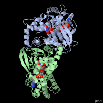
| Structural highlights
3int is a 2 chain structure with sequence from Klebsiella pneumoniae. Full crystallographic information is available from OCA. For a guided tour on the structure components use FirstGlance.
| | Ligands: | , ,
| | Related: | 1i8t, 2bi7, 2bi8, 1wam, 1v0j, 3gf4, 3inr |
| Gene: | glf, rfbD (Klebsiella pneumoniae) |
| Activity: | UDP-galactopyranose mutase, with EC number 5.4.99.9 |
| Resources: | FirstGlance, OCA, RCSB, PDBsum |
Evolutionary Conservation
Check, as determined by ConSurfDB. You may read the explanation of the method and the full data available from ConSurf.
Publication Abstract from PubMed
The flavoenzyme uridine 5'-diphosphate galactopyranose mutase (UGM or Glf) catalyzes the interconversion of UDP-galactopyranose and UDP-galactofuranose. The latter is a key building block for cell wall construction in numerous pathogens, including Mycobacterium tuberculosis. Mechanistic studies of UGM suggested a novel role for the flavin, and we previously provided evidence that the catalytic mechanism proceeds through a covalent flavin-galactose iminium. Here, we describe 2.3 and 2.5 A resolution X-ray crystal structures of the substrate-bound enzyme in oxidized and reduced forms, respectively. In the latter, C1 of the substrate is 3.6 A from the nucleophilic flavin N5 position. This orientation is consistent with covalent catalysis by flavin.
X-ray Crystallography Reveals a Reduced Substrate Complex of UDP-Galactopyranose Mutase Poised for Covalent Catalysis by Flavin .,Gruber TD, Westler WM, Kiessling LL, Forest KT Biochemistry. 2009 Sep 10. PMID:19719175[1]
From MEDLINE®/PubMed®, a database of the U.S. National Library of Medicine.
See Also
References
- ↑ Gruber TD, Westler WM, Kiessling LL, Forest KT. X-ray Crystallography Reveals a Reduced Substrate Complex of UDP-Galactopyranose Mutase Poised for Covalent Catalysis by Flavin . Biochemistry. 2009 Sep 10. PMID:19719175 doi:10.1021/bi901437v
|