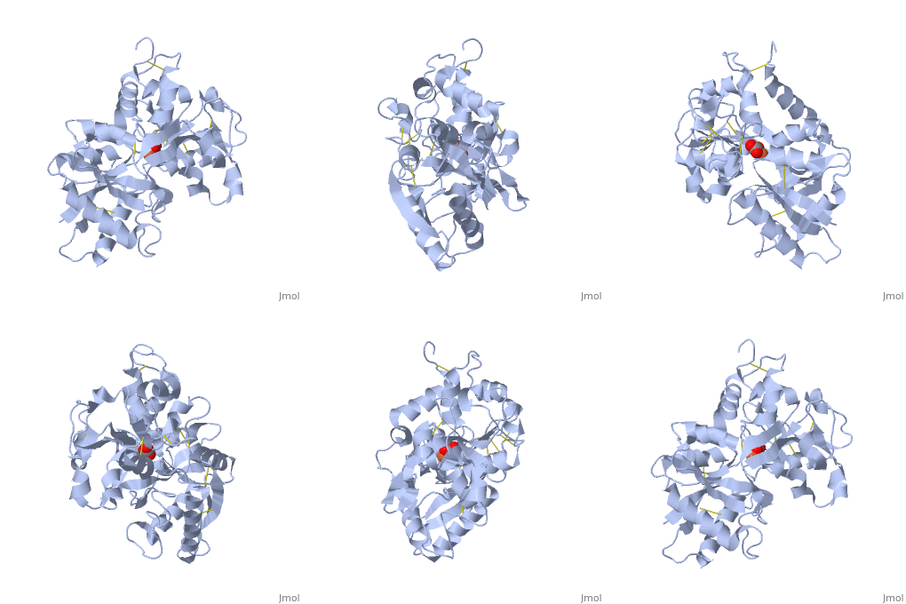
1dtg
From Proteopedia
| |||||||||
| 1dtg, resolution 2.40Å () | |||||||||
|---|---|---|---|---|---|---|---|---|---|
| Ligands: | , | ||||||||
| |||||||||
| |||||||||
| Resources: | FirstGlance, OCA, RCSB, PDBsum | ||||||||
| Coordinates: | save as pdb, mmCIF, xml | ||||||||
Contents |
HUMAN TRANSFERRIN N-LOBE MUTANT H249E
Template:ABSTRACT PUBMED 10684598
Disease
[TRFE_HUMAN] Defects in TF are the cause of atransferrinemia (ATRAF) [MIM:209300]. Atransferrinemia is rare autosomal recessive disorder characterized by iron overload and hypochromic anemia.[1][2]
Function
[TRFE_HUMAN] Transferrins are iron binding transport proteins which can bind two Fe(3+) ions in association with the binding of an anion, usually bicarbonate. It is responsible for the transport of iron from sites of absorption and heme degradation to those of storage and utilization. Serum transferrin may also have a further role in stimulating cell proliferation.
About this Structure
1dtg is a 1 chain structure with sequence from Homo sapiens. Full crystallographic information is available from OCA.
See Also
- Cartoon backbone representation
- Secondary structure
- Sheets in Proteins
- Spacefilling representation
- Transferrin
Reference
- MacGillivray RT, Bewley MC, Smith CA, He QY, Mason AB, Woodworth RC, Baker EN. Mutation of the iron ligand His 249 to Glu in the N-lobe of human transferrin abolishes the dilysine "trigger" but does not significantly affect iron release. Biochemistry. 2000 Feb 15;39(6):1211-6. PMID:10684598
- ↑ Beutler E, Gelbart T, Lee P, Trevino R, Fernandez MA, Fairbanks VF. Molecular characterization of a case of atransferrinemia. Blood. 2000 Dec 15;96(13):4071-4. PMID:11110675
- ↑ Knisely AS, Gelbart T, Beutler E. Molecular characterization of a third case of human atransferrinemia. Blood. 2004 Oct 15;104(8):2607. PMID:15466165 doi:10.1182/blood-2004-05-1751


