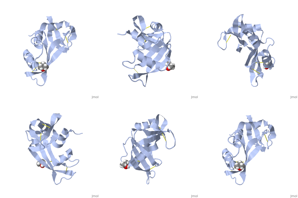
Structural highlights
Evolutionary Conservation
Check, as determined by ConSurfDB. You may read the explanation of the method and the full data available from ConSurf.
Publication Abstract from PubMed
The structure of phosphate-free bovine ribonuclease A has been refined at 1.26-A resolution by a restrained least-squares procedure to a final R factor of 0.15. X-ray diffraction data were collected with an electronic position-sensitive detector. The final model consists of all atoms in the polypeptide chain including hydrogens, 188 water sites with full or partial occupancy, and a single molecule of 2-methyl-2-propanol. Thirteen side chains were modeled with two alternate conformations. Major changes to the active site include the addition of two waters in the phosphate-binding pocket, disordering of Gln-11, and tilting of the imidazole ring of His-119. The structure of the protein and of the associated solvent was extensively compared with three other high-resolution, refined structures of this enzyme.
Structure of phosphate-free ribonuclease A refined at 1.26 A.,Wlodawer A, Svensson LA, Sjolin L, Gilliland GL Biochemistry. 1988 Apr 19;27(8):2705-17. PMID:3401445[1]
From MEDLINE®/PubMed®, a database of the U.S. National Library of Medicine.
See Also
References
- ↑ Wlodawer A, Svensson LA, Sjolin L, Gilliland GL. Structure of phosphate-free ribonuclease A refined at 1.26 A. Biochemistry. 1988 Apr 19;27(8):2705-17. PMID:3401445


