3hts
From Proteopedia
| |||||||
| , resolution 1.75Å | |||||||
|---|---|---|---|---|---|---|---|
| Ligands: | |||||||
| Coordinates: | save as pdb, mmCIF, xml | ||||||
HEAT SHOCK TRANSCRIPTION FACTOR/DNA COMPLEX
Overview
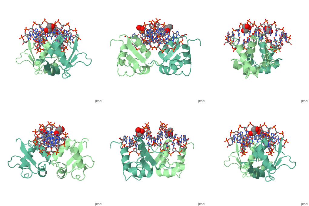
The 1.75 A crystal structure of the Kluyveromyces lactis heat shock transcription factor (HSF) DNA-binding domain (DBD) complexed with DNA reveals a protein-DNA interface with few direct major groove contacts and a number of phosphate backbone contacts that are primarily water-mediated interactions. The DBD, a 'winged' helix-turn-helix protein, displays a novel mode of binding in that the 'wing' does not contact DNA like all others of that class. Instead, the monomeric DBD, which crystallized as a symmetric dimer to a pair of nGAAn inverted repeats, uses the 'wing' to form part of the protein-protein contacts. This dimer interface is likely important for increasing the DNA-binding specificity and affinity of the trimeric form of HSF, as well as for increasing cooperativity between adjacent trimers.
About this Structure
3HTS is a Single protein structure of sequence from Kluyveromyces lactis. Full crystallographic information is available from OCA.
Reference
A new use for the 'wing' of the 'winged' helix-turn-helix motif in the HSF-DNA cocrystal., Littlefield O, Nelson HC, Nat Struct Biol. 1999 May;6(5):464-70. PMID:10331875
Page seeded by OCA on Thu Mar 20 19:05:21 2008

