This old version of Proteopedia is provided for student assignments while the new version is undergoing repairs. Content and edits done in this old version of Proteopedia after March 1, 2026 will eventually be lost when it is retired in about June of 2026.
Apply for new accounts at the new Proteopedia. Your logins will work in both the old and new versions.
Image:PRPc.png
From Proteopedia
Size of this preview: 752 × 600 pixels
Full resolution (770 × 614 pixel, file size: 357 KB, MIME type: image/png)
Summary
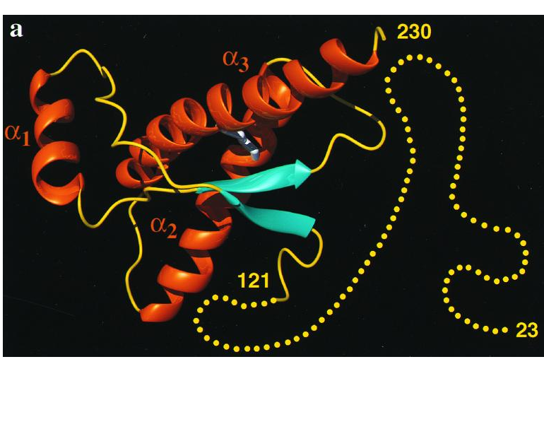
Cartoon of the three-dimensional structure of the intact human prion protein, hPrP(23–230). The helices are orange, theb-strands cyan, the segments with nonregular secondary structure within the C-terminal domain yellow, and the flexibly disordered ‘‘tail’’ of residues 23–121 is represented by yellow dots.
This image was taken with premission from Zahn, R., Liu, A., Luhrs, T., Riek, R., von Schroetter, C., Lopez Garcia, F., Billeter, M., Calzolai, L., Wider, G. Wuthrich, K. (2000). NMR solution structure of the human prion protein. The Proceedings of the National Academy of Sciences U.S.A., 97(1), 145-150.
Licensing
{{subst:Permission from license selector}}
File history
Click on a date/time to view the file as it appeared at that time.
| Date/Time | User | Dimensions | File size | Comment | |
|---|---|---|---|---|---|
| (current) | 07:29, 19 January 2015 | Nitzan Cohen Amin (Talk | contribs) | 770×614 | 357 KB | This image was taken with premission from Zahn, R., Liu, A., Luhrs, T., Riek, R., von Schroetter, C., Lopez Garcia, F., . . . Wuthrich, K. (2000). NMR solution structure of the human prion protein. The Proceedings of the National Academy of Sciences U.S.A |
- Edit this file using an external application
See the setup instructions for more information.
Links
The following pages link to this file:
