Classification
EC 3.4.24.34
This classification means that this enzyme:
- is an hydrolase: it hydrolyzes covalent bonds
- is an endopeptidase: cleaves peptide bond
- cleves interstitial collagens in the triple helical domain
The difference between this classification and EC 3.4.24.7 is that this enzyme cleaves type III collagen more slowly than type I.
To see
[1]
[2]
[2]
[3]
[4] pockets
[5]
Structure
Domains
Thanks to X-ray crystallography, the structure of 2OY4 has been solved with 1,7 Å resolution. This enzyme consists of and .
Pocket interaction
Ca2+
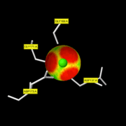
The active site of the enzyme is composed by two Gly residues (169 and 171) next to two Asp residues (137 and 173).
This enzyme binds 3 CA ions per subunit.
maintain a Zn2+ atom coordinated with three water molecules. One of them is as well bound to the Glu residue thanks to a hydrogen bond. The second Zn2+ atom is not involved in the active site. At first, the Gly 206 residue of the substrate binds the active site thanks to the Zn2+ atom. When it binds it takes the place of unstable water molecules and establishes stabilizing interactions with the active site thanks to its C terminal part. Then, the Ala 182 residue of the enzyme makes a hydrogen bond with the NH group of the substrate: this allows the substrate to enter the cavity of the catalytic site. The rest of the protein is stabilized by 4 hydrogen bonds with the amino acid located in the cavity.
Zn2+
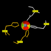
The active site of the enzyme is composed by one Asp residue (149) next to three His residues (147, 162 and 175).
This enzyme binds 2ZN ions per subunit.
maintain a Zn2+ atom coordinated with three water molecules. One of them is as well bound to the Glu residue thanks to a hydrogen bond. The second Zn2+ atom is not involved in the active site. At first, the Gly 206 residue of the substrate binds the active site thanks to the Zn2+ atom. When it binds it takes the place of unstable water molecules and establishes stabilizing interactions with the active site thanks to its C terminal part. Then, the Ala 182 residue of the enzyme makes a hydrogen bond with the NH group of the substrate: this allows the substrate to enter the cavity of the catalytic site. The rest of the protein is stabilized by 4 hydrogen bonds with the amino acid located in the cavity.
The conserved cysteine present in the cysteine-switch motif (89-96) binds the catalytic zinc ion, thus inhibiting the enzyme. The dissociation of the cysteine from the zinc ion upon the activation-peptide release activates the enzyme.
Catalytic domain
It seems to have 163 residues involved in the catalytic domain: Met80-Gly242.
(http://www.ncbi.nlm.nih.gov/pmc/articles/PMC394940/?page=1). The catalytic zinc ion is situated at the bottom of the active-site. The other zinc ion and the two calcium ions are packed against the top of the beta sheet and presumably function to stabilize the catalytic domain. The polypeptide folding and in particular the zinc environment of the collagenase catalytic domain bear a close ressemblance to the astacins and the snake venom metalloproteinases.
Function
Like all the MMPs, MMP8 is secreted as inactive proproteins and then activated after a cleavage by extracellular proteinases. Indeed, it can't be activated without removal of the .
cleave the helical collagen molecule at a single Gly-Ile/Leu bond in each alpha-chain of the molecule. The cleavage generates fragments that spontaneously lose their helical conformation, denature to gelatin, and become soluble. The gelatin is then susceptible to attack by gelatinases and other proteases[3]
metalloendopeptidase activity = mechanism in which water acts as a nucleophile, one or two metal ions hold the water molecule in place, and charged amino acid side chains are ligands for the metal ions.[4]
Disease
Overexpression of MMP8, or inadequate control by TIMPs, can be associated with a lot of pathological conditions: psoriasis, sclerosis, osteoarthritis, rheumatoid arthritis, osteoporosis, Alzheimer's disease, tumor growh and metastasis.[5]
Relevance
Structural highlights
This is a sample scene created with SAT to by Group, and another to make of the protein. You can make your own scenes on SAT starting from scratch or loading and editing one of these sample scenes.


