This old version of Proteopedia is provided for student assignments while the new version is undergoing repairs. Content and edits done in this old version of Proteopedia after March 1, 2026 will eventually be lost when it is retired in about June of 2026.
Apply for new accounts at the new Proteopedia. Your logins will work in both the old and new versions.
Sandbox HEC
From Proteopedia
1dgb
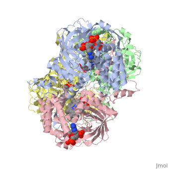
Human Erythrocyte CatalaseFunction1DGB is used to protect hemoglobin by removing hydrogen peroxide generated from erythrocytes. Human catalase is a heme-containing enzyme whose primary function is to break down hydrogen peroxide into two molecules of water and one molecule of oxygen. Human catalase plays a major part in the defense against oxidative damage and inactivation of hemoglobin by removing half of the hydrogen peroxide formed by human erythrocytes [1] . Hydrogen peroxide is a byproduct of normal cellular respiration, but is toxic at high concentrations. If catalase does not break down hydrogen peroxide broken, it gets converted into reactive oxygen species and can damage DNA, proteins, and cell membranes (Source-goth (3)). Human catalase enzyme has been noted as an important factor in inflammation, mutagenesis, prevention of apoptosis, and stimulation of tumors. During a normal catalytic cycle hydrogen peroxide is the source of both oxidative and reductive potential. NADPH has been known to also bind to human catalase, however it does not serve as the oxidative or reductive potential source (putnam).
Structure and MechanismHuman erythrocyte catalase is a negatively charged heme-containing monofunctional tetrameric enzyme prevalent among aerobic organisms [2][3] Cite error: Invalid The catalase fold, a stereoscopic alignment of the clade 3 subunits, contains an eight-sheeted anti-parallel beta-barrel domain linked to a six alpha-helical domain via a lengthy protein sequence. Residues within β1-β4 contribute to the heme variant, while monomers within β5-β8 establish the NADPH binding site (Diaz, Loewen, Fita, & Carpena, 2012). The positioning of the heme is determined by the proximal aromatic pyrrole compounds ; in human erythrocyte catalase, catalytic His75 is positioned above pyrrole ring III, further producing a His-III orientation and heme-b variant. The NADPH binding site is located at the β,α-domain junction (Alfonso-Prietro, Vidossich, & Rovira, 2012; Diaz, Loewen, Fita, & Carpena, 2012). When the NADPH molecule is bound, a right-handed clockwise helical formation is produced. In human erythrocyte catalase, only two of the four subunits allow for NADPH binding [6](Kodydková, Vávrová, Kocík, & Zák, A., 2014; Diaz, Loewen, Fita, & Carpena, 2012). The active site contains a negatively charged tyrosine and a positively charged histidine situated, respectively, proximal and distal to the heme group. The histidine is responsible for the formation of Compound I during the first step of the catalase mechanism (Alfonso-Prietro, Vidossich, & Rovira, 2012). The deeply buried heme group is connected to the protein surface by a primary channel which provides a transport pathway for the hydrogen peroxide substrate (Diaz, Loewen, Fita, & Carpena, 2012). The transportation of hydrogen peroxide through the main channel is regulated by electrical dipole interactions between the hydrogen peroxide and the hydrophobic portion of the channel containing negatively charged aspartate and positively charged iron from the heme (Lennicke et al., 2015; Diaz, Loewen, Fita, & Carpena, 2012; Halliwell, Clement, & Long, 2000). Additionally, less significant lateral channels allow products to leave the heme pocket (Diaz, Loewen, Fita, & Carpena, 2012). Human erythrocyte catalase is not evenly distributed throughout the body due to restricted endothelium passageways; this allows for a controlled and localized dissemination of the protein (Nishikawa, Hashida, & Takakura, 2009).
Disease and DisordersThere are 12 known mutations in the human erythrocyte catalase gene that have been found to cause acatalasemia (5). Acatalasemia is an autosomal recessive condition in which human erythrocyte catalase levels are very low. Most people are asymptomatic and are diagnosed because a family member is affected. However, although they are asymptomatic, they have an increased risk of chronic diseases. Acatalasemia can be correlated with ulcers and gangrene. When this occurs, the condition is known as Takahara disease. Ulcers and gangrene can result from high levels of hydrogen peroxide that is normally produced from bacteria. Mutations in the human erythrocyte catalase gene tend to reduce the activity of human erythrocyte catalase (1) to less than 10% of its normal activity thus reducing the enzymes ability to degrade hydrogen peroxide and causing a build-up of hydrogen peroxide. This build-up in turn causes ulcers and gangrene. A similar condition to acatalasemia is hypocatalasemia, in which each cell of the human erythrocyte catalase only has one gene with a mutation, instead of both genes with a mutation. This single mutation cuts the activity of human erythrocyte catalase by about half. Similar to acatalasemia, this condition normally doesn’t cause health issues (2). Acatalasemia is also associated with type 2 diabetes mellitus, the most common form of diabetes. The build-up of hydrogen peroxide from the decrease in human erythrocyte catalase can damage beta cells in the pancreas. The pancreas releases insulin, which helps your body regulate your blood sugar level. However, the damaged beta cells cannot utilize the insulin as well as normal beta cells, which leads to type 2 diabetes mellitus. These defective beta cells are thought to be why people with acatalasemia have an increased risk for type 2 diabetes mellitus. A larger percentage of people with diabetes have acatalasemia than those with diabetes without acatalasemia. Those with acatalasemia also tend to develop diabetes at an earlier age (1). Common variations in the human erythrocyte gene and variations in the regions of DNA that help to regulate the gene’s activity may also lead to an increased risk of a person developing specific common, complex diseases such as hypertension, osteoporosis, and heart attack and stroke due to the elevated levels of cholesterol and other fats in the blood (1,3,4). However, not all people experience health problems when they have a loss of catalase activity and others do not have an identified mutation in the human erythrocyte catalase gene when they have a loss in catalase activity. The cause of both of these situations is unclear. Some researched hypothesize that the activity is also influenced by other genetic factors as well as environmental conditions (1,5).
References
| ||||||||||||