This old version of Proteopedia is provided for student assignments while the new version is undergoing repairs. Content and edits done in this old version of Proteopedia after March 1, 2026 will eventually be lost when it is retired in about June of 2026.
Apply for new accounts at the new Proteopedia. Your logins will work in both the old and new versions.
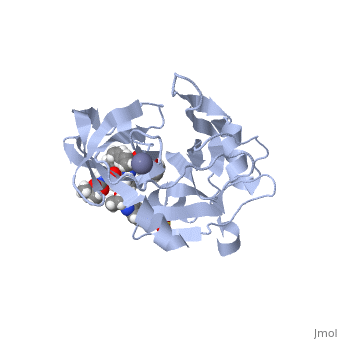
User:Megan Harris./Sandbox 1
From Proteopedia
| |||||||||||
References
- ↑ Hanson, R. M., Prilusky, J., Renjian, Z., Nakane, T. and Sussman, J. L. (2013), JSmol and the Next-Generation Web-Based Representation of 3D Molecular Structure as Applied to Proteopedia. Isr. J. Chem., 53:207-216. doi:http://dx.doi.org/10.1002/ijch.201300024
- ↑ Herraez A. Biomolecules in the computer: Jmol to the rescue. Biochem Mol Biol Educ. 2006 Jul;34(4):255-61. doi: 10.1002/bmb.2006.494034042644. PMID:21638687 doi:10.1002/bmb.2006.494034042644
- ↑ Coburn CA, Meinke PT, Chang W, Fandozzi CM, Graham DJ, Hu B, Huang Q, Kargman S, Kozlowski J, Liu R, McCauley JA, Nomeir AA, Soll RM, Vacca JP, Wang D, Wu H, Zhong B, Olsen DB, Ludmerer SW. Discovery of MK-8742: an HCV NS5A inhibitor with broad genotype activity. ChemMedChem. 2013 Dec;8(12):1930-40. doi: 10.1002/cmdc.201300343. Epub 2013 Oct, 14. PMID:24127258 doi:http://dx.doi.org/10.1002/cmdc.201300343
- ↑ National Center for Biotechnology Inforamtion. PubChem Compound Database; CID=71661251, https://pubchem.ncbi.nlm.nih.gov/compound/71661251#section=Chemical-and-Physical-Properties
- ↑ doi: https://dx.doi.org/http
- ↑ Fridell RA, Qiu D, Valera L, Wang C, Rose RE, Gao M. Distinct functions of NS5A in hepatitis C virus RNA replication uncovered by studies with the NS5A inhibitor BMS-790052. J Virol. 2011 Jul;85(14):7312-20. doi: 10.1128/JVI.00253-11. Epub 2011 May 18. PMID:21593143 doi:http://dx.doi.org/10.1128/JVI.00253-11
- ↑ Chevaliez, S.; Pawlotsky, J. M. Virology of hepatitis C virus infection. Best Pract. Res. Clin. Gastroenterol.2012, 26, 381-389.
- ↑ Petrescu, I. O.; Biciusca, V.; Taisescu, C. I.; Alexandru, D. O.; Taisescu, O.; Comanescu, M. V.; Petrescu, F.; Popescu, I. A.; Trasca, D. M.; ForTofoiu, M. C.; Silosi, C. A.; ForTofoiu, M. Histological factors that predict the liver fibrosis in patients with chronic hepatitis C. Rom. J. Morphol. Embryol. 2016, 57, 759-765.
